
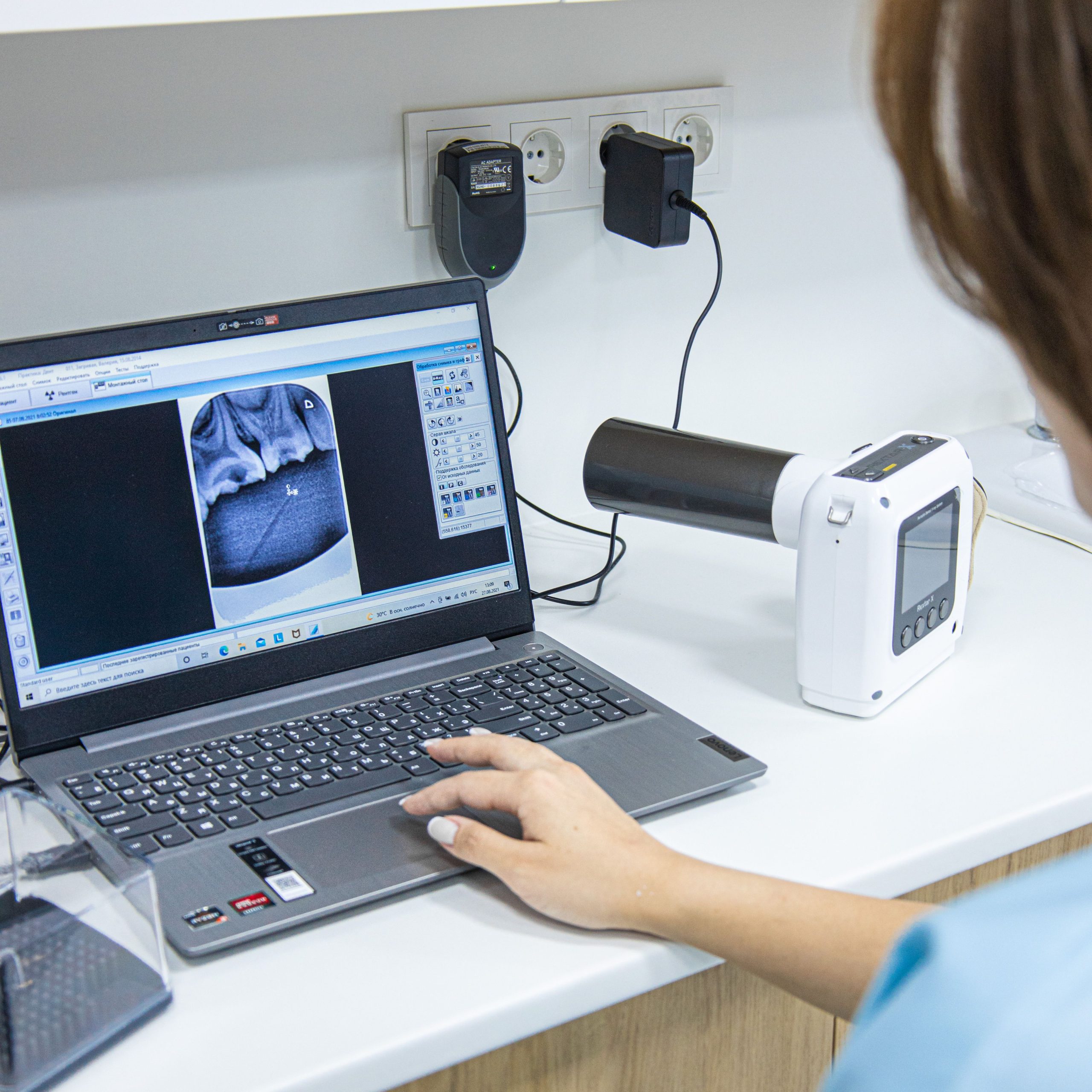
Every time a dentist asks a patient to stand in front of a panoramic X-ray machine and says, “Do not move,” a process far more complex than simply taking a picture is taking place. Within seconds, X-rays pass through the soft tissues of the mouth, and there, within the thin cells of the gingival mucosa, changes begin to occur at the molecular level.
This question prompted Dr. drg. Rurie Ratna Shantiningsih, MDSc, together with a research team from Universitas Gadjah Mada, to investigate what actually happens to the DNA of gingival cells following panoramic radiographic exposure. The resulting study, published in the September 2013 issue of Dental Journal (Majalah Kedokteran Gigi), revealed a significant correlation between two markers of genetic damage: micronuclei and 8-oxo-dG expression.
Two Markers, One Story of Damage
To understand the findings, it is important to identify the two main players.
First, micronuclei. These are small fragments of genetic material that become separated from the main cell nucleus as a result of chromosome damage. Their presence is more than a structural abnormality. Scientists use micronuclei as an early biomarker of carcinogenesis, signaling that chromosomes within cells have sustained significant damage.
Second, 8-hydroxy-2-deoxyguanosine, abbreviated as 8-oxo-dG. This compound forms when free radicals generated by X-ray ionization attack the guanine bases in DNA strands, producing what is known as a DNA adduct. Put simply, it is a “chemical fingerprint” of oxidative DNA damage.
Interestingly, these two markers had not previously been investigated simultaneously in the context of panoramic radiographic exposure.
Rabbits, X-rays, and Nine Days of Observation
The research team used 12 six-month-old male New Zealand rabbits as subjects. The animals were divided into four groups according to observation time: day 0 (immediately after exposure), day 3, day 6, and day 9.
Each rabbit was exposed to panoramic radiography using a Yoshida Panoura machine at 80 kVp and 8 mA for 12 seconds, producing a dose rate of 47 μSv. This dose is comparable to that commonly used in human clinical practice.
Swabs of the anterior mandibular gingival mucosa were collected before and after exposure to count micronuclei using a modified Feulgen-Rossenbeck staining method. Meanwhile, gingival tissue sections were analyzed by immunohistochemistry to detect 8-oxo-dG expression.
The results were notable. The number of micronuclei continued to increase over time, reaching a peak on day 9. This is consistent with previous research indicating that the turnover period of gingival epithelial cells ranges from 7 to 16 days, making days 9 and 10 an optimal period for detecting micronuclei.
In contrast, 8-oxo-dG expression showed the opposite pattern. Immediately after exposure, 8-oxo-dG expression was strongest and most widespread. However, over time, the staining intensity diminished and the area showing positive expression became increasingly smaller. By day 9, only approximately 10% of the area still showed positive expression.
A Negative Correlation with a Positive Biological Meaning
Pearson correlation analysis demonstrated a significant relationship between the increase in micronuclei and the decrease in 8-oxo-dG expression scores, with r = −0.658 and p = 0.020. Rather than being confusing, this negative correlation makes biological sense.
“The appearance of micronuclei in gingival epithelium can be detected after cells undergo exfoliation several days after exposure, while the expression of the DNA adduct represented by 8-oxo-dG decreases due to the body's physiological ability to eliminate the damage once exposure to the mutagen has ceased.”
The body, it turns out, does not remain passive. The base excision repair (BER) mechanism actively repairs single-base lesions in DNA, including 8-oxo-dG. While BER removes traces of oxidative damage, micronuclei that have already formed can persist for one or two cell division cycles. This explains why the two markers move in opposite directions over the same period.
Radiation Protection Is More Than a Formality
These findings have clinical implications that should not simply be overlooked. Even though a single panoramic radiographic exposure uses a standard clinical dose, genotoxic effects can still be detected in gingival cells. Although some of the resulting DNA damage can be repaired by the body's natural mechanisms, repeated exposure without clear clinical indications may create the potential for genetic mutations that could contribute to carcinogenesis.
The study also opens the door to further investigation into the use of antioxidants as radioprotective agents to reduce the increase in reactive oxygen species (ROS) caused by ionizing radiation. If proven effective, such a strategy could change how we approach the preparation and follow-up of patients undergoing repeated radiographic examinations.
Panoramic radiography remains a reliable and indispensable diagnostic tool in dentistry. Yet behind those clear images of the jawbone, a silent conversation is taking place within the cells—between radiation, DNA, and the body's defense mechanisms working largely unnoticed.
Source DOI: https://doi.org/10.20473/j.djmkg.v46.i3.p119-123
Authors: Anny Anggraini; drg. Achmad Zam Zam Aghasy, M.Kes.
Photo: Pexels