
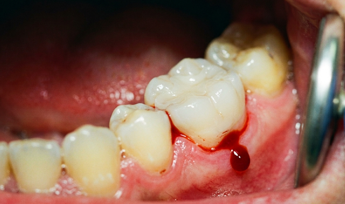
When dentists perform apical surgery, a surgical procedure used to save teeth that have failed conventional treatment, they face a very real challenge. The red fluid freely flowing through the surgical area is not merely an obstruction to visibility; it is also a direct threat to the filling material placed at the end of the tooth root. Prof. drg. Diatri Nari Ratih, M.Kes., Ph.D., Sp.KG(K), a researcher from the Department of Conservative Dentistry, Faculty of Dentistry, Universitas Gadjah Mada (FKG UGM), demonstrated this through a study published in the Journal of Medical and Biological Engineering in 2017. The study, conducted together with drg. Asri Riany Putri, Sp.KG., examined the extent to which blood contamination weakens the bond strength of Mineral Trioxide Aggregate (MTA), one of the leading filling materials currently used in endodontic surgery, and found that the choice of mixing liquid determines the severity of the damage.
Apical surgery is not a procedure undertaken lightly. Dentists generally recommend it only when conventional root canal treatment can no longer resolve the problem. Persistent infection, inflammation in the tissues surrounding the root tip, or a root canal that cannot be accessed from within the tooth crown can all be reasons for performing this procedure.
During apical surgery, the dentist opens the gum tissue, accesses the jawbone, and then removes the problematic tip of the root. The small cavity at the end of the resected root must then be tightly sealed with a special material known as a retrograde filling. The goal is straightforward: to prevent bacteria and irritants from entering the surrounding tissues through the root canal.
MTA is a primary choice for this purpose. This powder-based material, first introduced to dentistry in 1993, offers several advantages not shared by many other materials: good sealing ability, biocompatibility, support for tissue regeneration, and the ability to set even under moist conditions. However, one question had not been fully answered before this study: what happens to the bond strength of MTA when it comes into direct contact with blood?
To answer this question, Prof. Diatri and her team designed an experiment using 30 human mandibular premolar tooth samples. Each tooth was sectioned at the middle of the root to produce 3-mm-thick slices, and a hole with a standardized diameter of 1.3 mm was created in the center, resembling the cavity that would be filled with restorative material during an actual surgical procedure.
The 30 root slices were divided into three groups according to the liquid used to mix the MTA powder. The first group used sterile water, the standard liquid recommended by the manufacturer. The second group used a 2% lidocaine-based local anesthetic because, in clinical practice, when sterile water is unavailable, local anesthetic is often an easily accessible substitute. The third group used a 5% calcium chloride (CaCl₂) solution, an accelerator added with the expectation of reducing the setting time of MTA.
Each group was then divided into two subgroups. Some samples were contaminated with fresh human blood injected directly into the cavity before MTA placement, while the others were left uncontaminated. All specimens were then stored in a 37°C incubator for 72 hours to simulate human body conditions before being tested.
Testing was performed using the push-out method, in which MTA is pushed out of the cavity using a specialized instrument attached to a universal testing machine. The maximum force required to dislodge the MTA from the dentin walls was recorded and then converted into megapascals (MPa), the standard unit for measuring bond strength in dental-material research.
The results showed that blood contamination reduced MTA bond strength in every group without exception. However, the extent of the reduction varied depending on the mixing liquid used. In the groups without blood contamination, the three liquids produced relatively similar bond strengths: 5% CaCl₂ produced the highest value at 22.14 MPa, followed by local anesthetic at 21.03 MPa and sterile water at 19.15 MPa. Once blood was introduced, the gap widened dramatically. MTA mixed with 5% CaCl₂ remained the most resistant, with a bond strength of 13.48 MPa. MTA mixed with sterile water dropped to 7.55 MPa. Most concerningly, MTA mixed with local anesthetic retained a bond strength of only 5.29 MPa, representing a decrease of more than 75% from the uncontaminated condition.
These figures illustrate something clinically important. In oral surgery, a filling material that loses three-quarters of its bond strength because of blood contamination is vulnerable to dislodgement from its intended site. If this occurs, the entire purpose of the surgical procedure may be compromised.
Chemically, the study provides a plausible explanation for this phenomenon. MTA works by forming crystals during the setting process. These needle-shaped crystals, known as ettringite, play an important role in locking the entire mass of the material together, allowing it to become solid and adhere firmly to the dentin walls. Blood interferes with the formation of these crystals.
In addition, blood contains various cells and proteins, including albumin. These proteins can block the dentinal tubules—the microscopic channels within dentin—and fill the space between the MTA and the root wall. As a result, the biomineralization process that should normally occur, namely the formation of a hydroxyapatite layer that chemically binds MTA to dentin, is disrupted from the outset.
The reason local anesthetic produced the poorest results lies in the acidity of the liquid. Local anesthetics are acidic, and this acidity is known to increase the porosity of MTA after setting. The more porous a material becomes, the more easily it can dissolve and the weaker its mechanical strength becomes. The combination of the acidic nature of the local anesthetic and interference from blood contamination resulted in the lowest bond strength among all the groups tested.
By contrast, 5% CaCl₂ proved to be the most advantageous mixing liquid. The solution reduced the setting time of MTA to approximately 35 minutes, considerably faster than sterile water. With faster setting, the material has less time to become contaminated by continuously flowing blood. In addition, CaCl₂ increases the density of the MTA crystal structure and reduces porosity, making the material more resistant to forces attempting to dislodge it from the root wall.
This study is more than a laboratory exercise. Every day, in oral surgery clinics around the world, dentists face decisions such as: what liquid should be used to mix MTA when sterile water is unavailable? Or is the local anesthetic already on hand safe enough to use?
Prof. Diatri's findings provide a less encouraging answer for the latter practice. Although local anesthetic is sterile and readily available, using it as a mixing liquid for MTA—particularly in surgical procedures involving blood exposure—resulted in the lowest bond strength. This means that the risk of retrograde filling failure may increase.
On the other hand, using 5% CaCl₂ as both an accelerator and a mixing liquid provides a dual benefit: it accelerates setting and improves bond strength, even under blood-contaminated conditions. These findings support clinical consideration of preparing this solution as a more reliable alternative.
The study also emphasizes that dental materials cannot be evaluated solely under ideal laboratory conditions. Real-world conditions, including the presence of blood, oral fluids, and various other contaminants, must also be taken into account when establishing clinical procedure standards.
Under stereomicroscopic examination, most samples demonstrated a mixed pattern of adhesive and cohesive failure. This means that some MTA material remained attached to the dentin wall, while other portions separated together with the material itself. This finding indicates that after 72 hours, the bond between MTA and dentin had begun to develop from a purely mechanical bond into a more complex chemical bond.
Author: drg. Achmad Zam Zam Aghasy, M.Kes., Annisa Dwi Noviyanti
Photo: FreePik
Source DOI: https://doi.org/10.1007/s40846-016-0199-8