
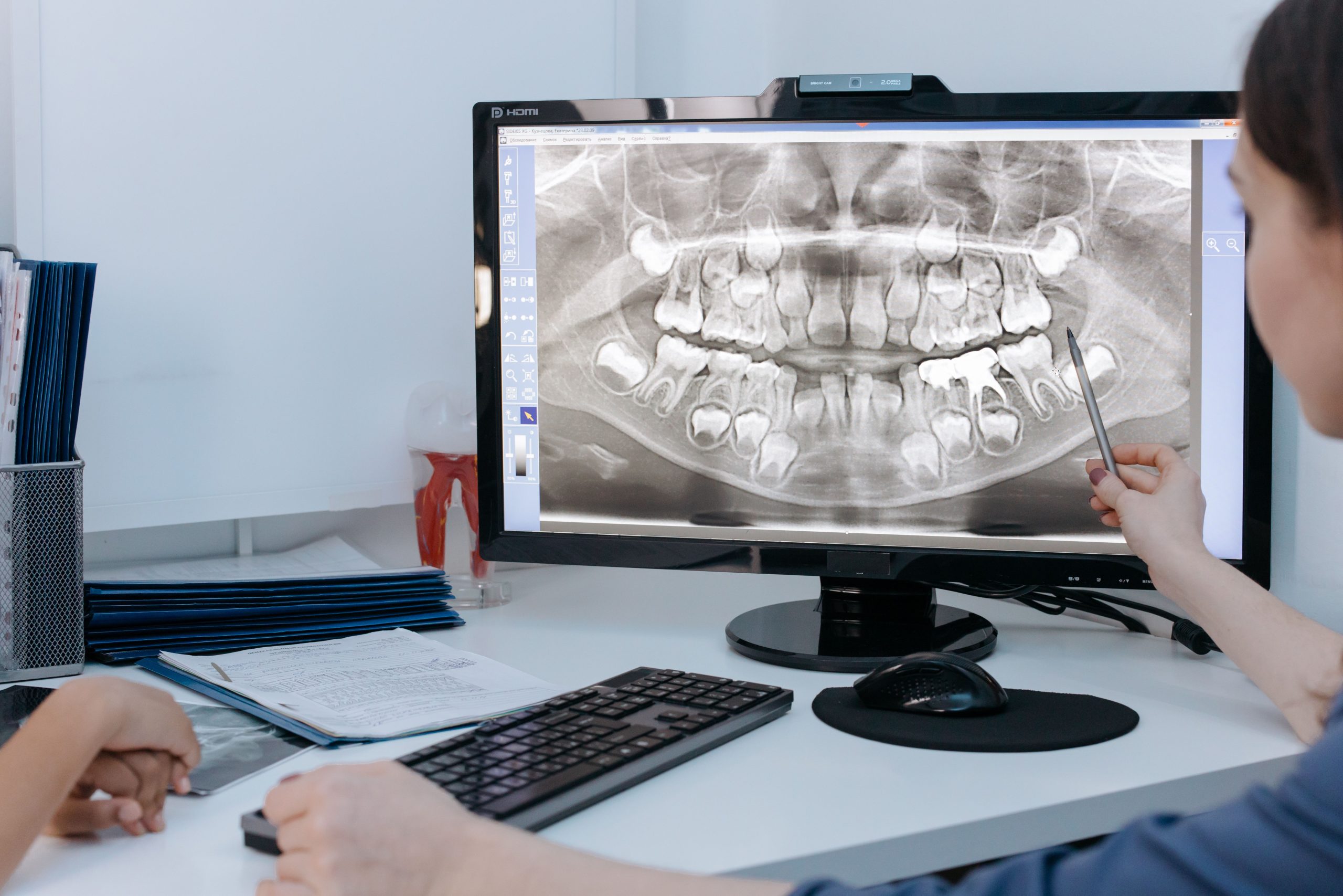
One in three women will spend roughly one-third of her life in menopause. It is not simply a matter of hormonal changes, but also of bones silently becoming weaker, more fragile, and increasingly vulnerable to fractures. The question is: can these changes be detected early, before significant damage occurs?
drg. Silviana Farrah Diba, Sp. RKG. Subsp. RP (K), from the Department of Dentomaxillofacial Radiology, Faculty of Dentistry, Universitas Gadjah Mada, sought to answer this question from an unusual perspective: through Cone Beam Computed Tomography (CBCT) images of the mandible. Together with a team from Universitas Padjadjaran, she investigated whether the trabecular microstructure of the mandibular bone in menopausal women could reflect levels of Beta-CrossLaps (β-CTx), a biochemical marker of bone resorption recommended by the International Osteoporosis Foundation.
The findings, published in the Journal of International Dental and Medical Research in 2020, offer a new perspective on the potential role of dental radiography in the early detection of osteoporosis.
From Blood to Images: Searching for a Meaningful Correlation
β-CTx is a fragment of type I collagen released into the bloodstream when bone undergoes resorption. The higher the level, the more active the bone degradation process. Traditionally, β-CTx is measured through clinical laboratory testing using a blood sample collected after a 12-hour fast and analyzed using the Electrochemiluminescence Immunoassay (ECLIA) method.
Meanwhile, CBCT has long been used in dental clinics for diagnosis, implant planning, and jaw evaluation. The technology can produce three-dimensional images at a lower cost than conventional CT. Interestingly, CBCT can also be used to analyze trabecular bone microstructure parameters, something that previously could only be performed using micro-CT or HR-pQCT, which are less practical for clinical patients.
In this study, 17 menopausal women aged 55 to 79 years underwent mandibular CBCT examinations. From each radiograph, a 3 × 3 mm Region of Interest (ROI) was selected in the trabecular area around the mental foramen, producing a total of 34 ROIs analyzed using BoneJ software. Three trabecular parameters were measured: bone volume fraction (BV/TV), trabecular thickness (Tb.Th), and trabecular separation (Tb.Sp).
Trabecular Separation: The Most Informative Marker
Of the three parameters examined, only Tb.Sp showed a significant correlation with β-CTx.
“Tb.Sp is the most sensitive microstructural parameter for detecting bone resorption or bone loss using CBCT in menopausal women in relation to β-CTx.”
The positive correlation found (r = 0.452, p < 0.05) means that the wider the distance between trabeculae, the higher the β-CTx concentration in the blood. Biologically, this makes sense. Estrogen deficiency in postmenopausal women triggers increased bone resorption, gradually altering trabecular architecture from plate-like structures into thin, rod-like structures with reduced connectivity. As a result, the spaces between trabeculae become wider.
By contrast, BV/TV and Tb.Th did not show significant correlations with β-CTx, although their trends were biologically plausible. BV/TV showed a weak negative correlation (r = −0.252), indicating that higher β-CTx levels tended to be associated with a lower percentage of bone volume. Meanwhile, Tb.Th showed a weak positive correlation (r = 0.227), a finding explained by a technical effect: the relatively large voxel size of CBCT makes thin trabeculae difficult to detect accurately, causing Tb.Th measurements to be overestimated because of the partial volume effect.
Great Potential, with Important Caveats
These findings open up an intriguing possibility: dentists, who routinely work with jaw radiographs, could potentially become part of the front line in early screening for bone loss among menopausal women. Existing equipment in dental clinics could be used for purposes extending beyond dental and jaw diagnosis alone.
However, the study also warrants caution. Its small sample size—only 17 participants—along with the small ROI size and uncontrolled nutritional factors, represents important limitations acknowledged by the researchers. The CBCT voxel size also affects the accuracy of Tb.Th and Tb.Sp measurements, meaning that the findings should be interpreted in light of the technical specifications of the imaging equipment.
Nevertheless, the direction of the research is clear: integrating routine radiographic examinations in dental clinics with clinical laboratory markers could provide a more holistic and affordable approach to assessing bone quality, particularly among the growing population of older women.
Bones may not speak. But with the right images, they can tell us far more than we might expect.
Source DOI: https://doi.org/10.5281/zenodo.20719795
Authors: Anny Anggraini; drg. Achmad Zam Zam Aghasy, M.Kes.
Photo: Pexels