
A scientific review from the Department of Conservative Dentistry, Faculty of Dentistry, Universitas Gadjah Mada (UGM), has revealed an interesting finding. A combination of two different materials, platelet-rich fibrin (PRF) and mineral trioxide aggregate (MTA), may represent a breakthrough in the treatment of immature permanent teeth with open apices resulting from trauma or infection. The review was published in the international journal Biomaterials Translational in 2026 and was authored by drg. Rahmadani Puspitasari, M.DSc., together with several colleagues from UGM and Universitas Airlangga, with Dr. drg. Tunjung Nugraheni, M.Kes., Sp.KG.(K), serving as the corresponding author.
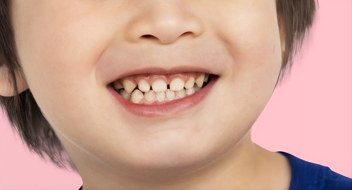
Imagine a 10-year-old child falling from a bicycle and severely hitting a tooth. The injury does not merely damage the crown of the tooth; it can also damage the blood vessels inside the root and cause pulp necrosis before the root has finished developing.
This condition is known as an immature tooth with an open apex. Its root is short, its walls are thin like an eggshell, and its root tip remains widely open. Without a closed apex, dentists do not have a sufficient “floor” on which to place root canal filling material. The risk of the material extruding into the tissues surrounding the root is therefore very high and may trigger serious inflammation.
For decades, calcium hydroxide has been the primary material used to close these open apices through a procedure known as apexification. However, the process is time-consuming, requires multiple visits, and can make the tooth root increasingly fragile. Then came MTA, a bioceramic material capable of forming an apical plug in a single visit, with significantly better sealing ability. The problem is that MTA acts like a static plug. It seals the apex but does not actively stimulate tissue recovery and further development.
This is where PRF enters as a promising complement. PRF is a biomaterial prepared from the patient's own blood. The procedure is relatively simple: blood is drawn from a vein, centrifuged for several minutes, and the middle layer that forms—a fibrin clot rich in platelets and white blood cells—is collected and used directly.
No additional chemicals are required. There is no risk of an immune reaction to a foreign material. PRF contains various natural growth factors, including TGF-β, VEGF, and PDGF, which promote cell growth, new blood vessel formation, and tissue regeneration. These factors are released gradually over 7 to 28 days after placement, providing sufficient support during the initial healing process.
In the context of apexification, PRF serves as an internal matrix. It is first placed at the root apex, forming a biological “cushion” that prevents MTA from extruding into the tissues outside the root. Once the PRF is in place, MTA is placed over it as a mechanically strong apical plug.
“The combination of PRF and MTA should not be viewed merely as a biological variation of conventional apexification, but rather as a translational strategy selected for teeth with open apices in which both apical control and biological support are clinically required.” — Puspitasari et al., Biomaterials Translational, 2026
The review analyzed 13 studies published between 2013 and 2024, retrieved from three major scientific databases: ScienceDirect, PubMed, and Scopus. Most were case reports, with only one clinical trial among them.
The results were relatively consistent: patients treated with the PRF-MTA combination generally showed favorable outcomes. Pain symptoms resolved, periapical lesions decreased radiographically, and in some cases, the apex gradually narrowed and the root walls became thicker. One clinical trial involving 27 patients even reported a 100% success rate after six months of follow-up.
From a biological perspective, the research explains why the combination makes scientific sense. MTA creates an alkaline environment with a pH above 12 that is antibacterial while also promoting the deposition of hydroxyapatite crystals, a natural mineral component of bone and cementum. PRF, meanwhile, provides a three-dimensional scaffold rich in growth factors that supports progenitor cell migration, new blood vessel formation, and tissue maturation.
The two materials work at different levels while complementing each other: MTA builds a mineral “wall,” while PRF prepares a fertile “living environment” in which cells can migrate and develop.
The review also discusses various types of PRF, including L-PRF, A-PRF, I-PRF, and Titanium PRF. Each has different characteristics in terms of consistency, cellular content, and growth-factor release rates. The choice of PRF type should be tailored to the clinical situation. A very wide apex may require PRF in the form of a dense membrane capable of supporting the MTA, whereas other situations may be better suited to a more fluid form of PRF that can spread more easily.
The researchers were cautious in drawing conclusions. Although the results appear positive, most of the evidence still comes from individual case reports involving small numbers of patients and varying protocols. Follow-up periods also varied considerably, ranging from just one week to two years.
There are still no large studies directly comparing whether the PRF-MTA combination is truly superior to MTA alone. Questions regarding how long the protective effect lasts, how the alkaline environment created by MTA affects the activity of PRF growth factors, and what standardized protocol could be consistently reproduced across clinical settings all await answers from larger, well-controlled studies. What is already clear is that this approach is biologically plausible, clinically promising, and worthy of continued investigation. For children whose teeth are injured before their roots have fully developed, the PRF-MTA combination may be more than simply a way to close a hole at the root tip—it may give the tooth a second chance to survive for many years.
Author: drg. Achmad Zam Zam Aghasy, M.Kes., Annisa Dwi Noviyanti
Photo: FreePik
Source DOI: https://doi.org/10.12336/bmt.25.00202