
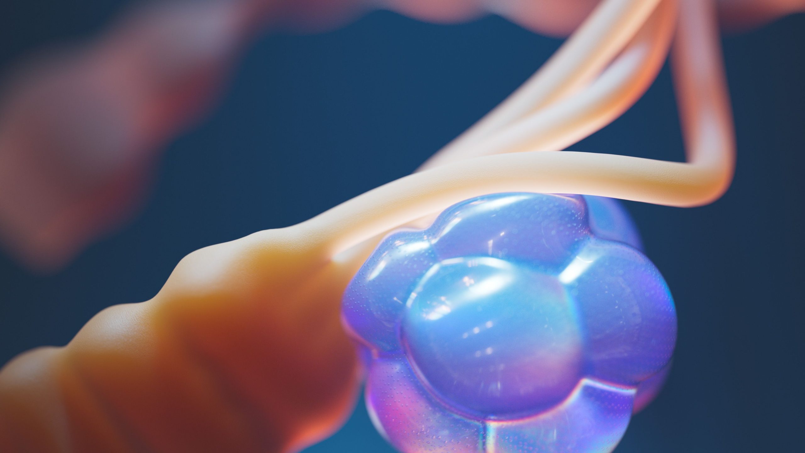
Imagine “aging” cells becoming architects that construct new tissue inside the womb. The finding may sound paradoxical, but this is what a recent study involving drg. Finsa Tisna Sari, M.Biomed., Ph.D., and a research team from Niigata University, Japan, has revealed. Published in the Journal of Oral Biosciences in August 2025, their study offers a new perspective on how palatal rugae—the patterned ridges on the hard palate—form during the earliest stages of life.
Tiny Ridges on the Palate That Hold a Major Secret
Palatal rugae are ridged structures found in all mammals, including humans and mice. They have traditionally been known to help break down food and guide tongue positioning during speech. However, behind their seemingly simple functions lies a complex and highly organized molecular mechanism governing their formation.
In mice, rugae begin to form on embryonic day (E) 12, beginning with localized thickening of the palatal epithelium known as a placode. Unlike the surrounding cells, cells within the placode do not proliferate. This has raised a long-standing question: what causes these cells to stop dividing with such precision?
The researchers suspected that the answer lay in senescence, a condition in which cells undergo permanent cell-cycle arrest. This condition is commonly associated with aging or disease rather than with the developmental construction of organs in embryos.
Evidence Emerges from Blue Staining
To detect senescence, the researchers used a classic marker called senescence-associated β-galactosidase (SA-β-gal). When the tissue was stained, senescent cells appeared blue. The results were surprising.
SA-β-gal activity was consistently detected in rugae placodes 1 through 4 and ruga 8 between embryonic days E12 and E14. As new rugae formed, the blue signal gradually disappeared, and by E16, when the ridged rugae structures were nearly complete, no SA-β-gal signal remained throughout the palatal tissue.
At the same time that the senescence signal disappeared, expression of the macrophage marker F4/80 emerged in the mesenchymal tissue beneath the placodes. This suggests that senescent cells are “cleared” by macrophages after completing their task, similar to what occurs during the development of embryonic limbs and kidneys.
Two other senescence markers, IL-6 and p21, were also found to be expressed in the rugae placodes during the same developmental stage as SA-β-gal activity. The number of p21-positive cells in the third ruga placode was statistically higher than that in the surrounding interplacodal epithelium, further supporting the idea that senescence is specifically localized to the regions where rugae develop.
An Unexpected Pathway: p21 Is Not the Master Regulator
Senescence during organ development has traditionally been understood to be activated through a p53-independent pathway involving p21. The researchers therefore conditionally deleted the p21 gene in mouse epithelium using the K14Cre system, generating p21^fl/fl;K14Cre mutant mice.
The result was both surprising and puzzling: SA-β-gal remained detectable in the mutant mice on E14, even in the absence of p21. This means that senescence in palatal rugae is induced through a p21-independent pathway, unlike the mechanisms identified in embryonic limbs and mesonephros.
“SA-β-gal activity persisted in mice with epithelial conditional deletion of p21. Furthermore, we successfully reduced SA-β-gal activity using a senolytic drug.” — Finsa Tisna Sari et al., Journal of Oral Biosciences, 2025
To verify that the detected cells were indeed genuine senescent cells, the team used a combination of senolytic drugs, Dasatinib and Quercetin, which were injected into pregnant mice from E11 to E13. In 10 of the 17 embryos examined, SA-β-gal activity was significantly reduced. Furthermore, no signs of apoptosis were found in the embryos treated with the drugs, indicating that the reduction in the signal was not caused by drug toxicity.
Interestingly, despite the successful elimination of senescent cells, the rugae pattern still developed normally, with no detectable abnormalities. This finding suggests that embryonic organogenesis possesses remarkable resilience: when one pathway is disrupted, other mechanisms can compensate.
When “Aging” Means Building
This study expands our understanding of senescence as a phenomenon that is not merely a sign of damage or aging, but also a programmed physiological mechanism involved in organ development. Embryonic senescence has previously been identified in the limbs, tail, otic vesicles, hindbrain, neural tube, mesonephros, and pharyngeal arches. Palatal rugae now join that list.
The implications extend beyond dentistry. Understanding how senescent cells are activated, function, and subsequently cleared during organ development could help researchers investigate the causes of congenital malformations, including palatal abnormalities, which remain a clinical challenge worldwide.
The specific molecular pathway that induces senescence in palatal rugae without the involvement of p21 remains an open question. And that is where this study leaves its mark—not as the final answer, but as the opening of a door that has only just been discovered.
Source DOI: https://doi.org/10.1016/j.job.2025.100688
Authors: Nanda Ayu, drg. Achmad Zam Zam Aghasy, M.Kes.
Photo: Pexels