
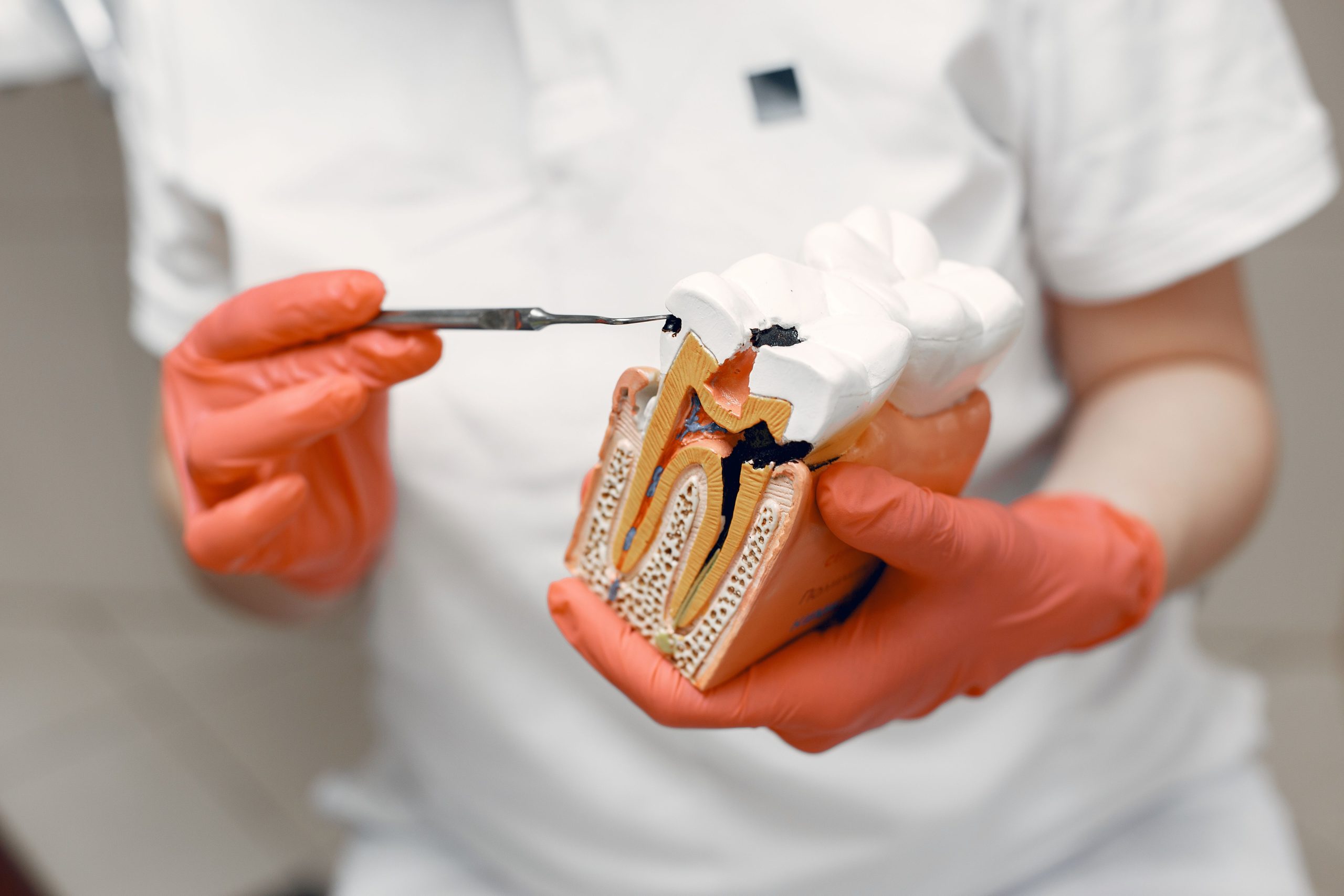
Imagine a defensive wall standing neatly at the edge of the pulp chamber. A layer of tall cells lines up like a fence, protecting the living tissue inside the tooth from external threats. These are odontoblasts. And when caries begins to erode the tooth, it is this wall that collapses first.
Research conducted by Prof. drg. Tetiana Haniastuti, M.Kes., Ph.D., from the Department of Oral Biology, Faculty of Dentistry, Universitas Gadjah Mada, directly demonstrates how the invasion of cariogenic bacteria damages the odontoblast layer in human teeth, cell by cell, stage by stage. The results were published in Dental Journal (Majalah Kedokteran Gigi) in September 2011 and became an important reference for understanding the biological response of the pulp to caries.
The Tooth Looks Fine, but the Cells Inside Are Already on Alert
To understand the damage, we first need to know what is being attacked. Odontoblasts are the outermost cells of the pulp. They are not merely passive occupants; they are producers of dentin, the hard layer that protects the pulp from the outside. Because of their peripheral position, they are the first to “sense” the presence of cariogenic bacteria.
Prof. Tetiana used 20 third molars extracted from volunteers aged 20 to 40 years. Five teeth were in normal condition, while the other 15 had caries at various depths: five teeth with caries limited to the enamel, five with caries extending into the dentin, and five with caries that had already reached the pulp. Each specimen was processed using hematoxylin-eosin staining, Gram staining to identify bacteria, and nestin immunohistochemistry, a specific marker indicating whether odontoblasts were still alive and functioning.
The results were surprising in their simplicity. In normal teeth, the odontoblast layer stood upright, was neatly arranged, and showed a strong nestin-positive reaction. But even in caries limited to the enamel, where bacteria had not actually entered the dentin, the odontoblast layer already showed signs of mild disturbance. Vacuolization appeared, resembling small “bubbles” inside the cells, indicating that soluble bacterial products had begun to seep through the dentinal tubules.
When Bacteria Enter the Dentin, the Fence Begins to Break Down
When caries had penetrated the dentin, the picture changed dramatically. Bacteria stained with Gram staining were clearly visible inside the dentinal tubules in all specimens. And beneath the lesion, the odontoblast layer could no longer be identified as an intact layer.
What remained were individual cells scattered sparsely, with altered morphology: flattened and no longer possessing their tall columnar shape. Some still showed a nestin-positive reaction, indicating that they were alive, but they were no longer functioning as an organized protective layer. Predentin, the layer that should form as a result of odontoblast activity, was also absent from the area beneath the lesion.
“The invasion of cariogenic bacteria can cause odontoblast cell damage by affecting their morphology and vitality. Cell damage becomes more severe as bacterial invasion progresses deeper toward the pulp.” — Prof. drg. Tetiana Haniastuti, M.Kes., Ph.D.
Interestingly, in areas farther away from the lesion, the odontoblast layer remained intact and active. This indicates that the damage was localized and progressive rather than occurring simultaneously throughout the tissue. The body was still attempting to defend itself in areas that had not yet been reached by bacteria.
When the Pulp Is Exposed: The Point of No Return
In cases of caries involving the pulp, the condition is already very different. Bacteria have successfully penetrated into the pulp chamber. The odontoblast layer on the roof of the pulp chamber can no longer be recognized in its normal arrangement. Only a few individual flattened cells remain and still show nestin-positive reactions, while severe inflammation is marked by massive infiltration of PMN (polymorphonuclear) cells and mononuclear cells dominating the microscopic appearance.
One finding is particularly noteworthy: nestin expression actually increased in pulp cells surrounding the inflamed area. This is not an ordinary recovery signal. The researchers interpreted it as an attempt by the body to coordinate the differentiation of new cells, functioning like an emergency signal calling on the remaining pulp cells to transform into new odontoblasts. Unfortunately, when the infection has reached this extent, the signal often comes too late.
Caries Is Not Just a Hole in the Tooth
What makes Prof. Tetiana’s research important is not only its technical findings but also its clinical implications. Caries has often been understood as a surface problem: a hole that needs to be filled. However, this research shows that long before a dentist sees a deep cavity, cellular damage has already begun within the pulp.
Damaged odontoblasts can no longer produce reactive dentin to seal off the threat. Without new predentin, dentinal tubules become an unobstructed pathway for bacteria toward the center of the pulp. And once the pulp becomes infected, treatment options become sharply limited.
For clinicians, this provides a strong scientific argument for early detection and prompt intervention. For researchers, it opens deeper questions: is there a way to strengthen or regenerate the odontoblast layer before the damage becomes irreversible? The answer to that question may be the key to the next generation of pulp therapy.
DOI: —
Authors: Anny Anggraini, drg. Achmad Zam Zam Aghasy, M.Kes. Photo: Pexels
Photo: Pexels