
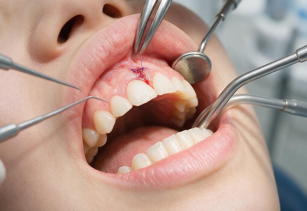
A lecturer and researcher from the Department of Conservative Dentistry, Faculty of Dentistry, Universitas Gadjah Mada (FKG UGM), drg. Margareta Rinastiti, M.Kes., Sp.KG(K)., Ph.D., has successfully formulated a new medical innovation to accelerate healing after gum surgery. Through in-depth research, she found that transplantation of human amniotic membrane, or the thin layer of the fetal membrane, can significantly stimulate the regeneration of gum tissue (gingiva), much faster than the natural healing process without wound coverage.
This valuable finding not only provides fresh hope for the field of dentistry but has also successfully brought its contribution to the global level through publication in the reputable international journal International Journal of Oral and Maxillofacial Surgery. This experiment opens significant opportunities for utilizing amniotic membranes, which have often been considered medical waste after childbirth, to be transformed into a “biological plaster” of the future that facilitates recovery in oral surgery patients.
Clinically, wounds in gum tissue following periodontal surgery or soft-tissue reconstruction are often a challenge in themselves. For a wound to close completely, the human body requires the role of cells called fibroblasts. These cells act as building units responsible for producing collagen and specialized proteins to patch and repair damaged tissue. Under normal conditions, fibroblasts in the mouth tend to remain passive. Once an injury occurs, these cells become activated, migrate, and begin to divide on the fourth or fifth day. Given that the oral cavity is a dynamic environment and vulnerable to friction from food and exposure to bacteria, the slow movement of fibroblasts often causes gum wounds to remain painful for days.
This is where the amniotic membrane acts as a tactical stimulus. The innermost layer of the fetal membrane is rich in growth factors or natural growth-promoting substances such as bFGF, EGF, and TGF-β. This series of active substances acts as a powerful molecular signal that drives fibroblast cells to migrate and function several times earlier than their normal biological rhythm. In addition, the membrane also contains laminin, a specialized substrate that facilitates the attachment of new gum epithelial cells and enables them to cover the wound area more rapidly.
To demonstrate the effectiveness of this innovation, drg. Margareta conducted a controlled experiment using an animal model in the laboratory. The study also involved strong cross-institutional and international collaboration, involving the Department of Anatomical Pathology, Faculty of Medicine, UGM; the Department of Pedodontics, FKG UGM; the Tissue Bank of Dr. Soetomo Regional General Hospital/Faculty of Medicine, Universitas Airlangga, Surabaya; and the Department of Oral Biology and Immunology, Universiti Sains Malaysia. The freeze-dried amniotic membrane was applied in five layers over the gum wounds of the samples and then carefully sutured to keep it stable before its development was evaluated from day 1 through day 14.
Histological observations under a light microscope showed that the inflammatory response was immediately controlled from the first day. This was demonstrated by a much lower number of inflammatory cells, thereby reducing swelling and pain. By the third day, new blood vessels, or the process of angiogenesis, had begun to form to supply nutrients to the new tissue. This rate surpassed that of the group without wound coverage, in which new blood vessels did not appear until the fifth day.
The peak of healing began to become clearly visible on the tenth day. At this stage, the number of fibroblast cells in the gums protected by the membrane reached its highest level, while the collagen fibers formed were not only abundant in quantity but also dense and very well organized, or mature. The recovery phase was finally completed earlier, on the fourteenth day, because the gums treated with the amniotic membrane had completed the tissue remodeling stage. This was in contrast to the natural-healing group, which on the fourteenth day was only beginning to produce new fibroblasts, indicating that its healing process was considerably behind.
In addition to being rich in growth-promoting substances, human amniotic membranes have another special advantage: a very low level of immune rejection. This occurs because the fetal membrane expresses immunoregulatory molecules that prevent the recipient’s body from recognizing it as a foreign threat, making it very safe for use across individuals.
For the general public and clinical practitioners, this preliminary research lays a very important scientific foundation. Postoperative gum wounds may appear minor, but for patients experiencing them, every day that can be cut from the recovery process so that they can return to eating, drinking, and speaking comfortably is certainly meaningful. Beginning with her master’s studies at UGM, this innovation in utilizing fetal membranes demonstrates that biomaterials-based research can provide real solutions to support healthy and prosperous lives in the community.
Author: drg. Achmad Zam Zam Aghasy, M.Kes., Annisa Dwi Noviyanti
Photo: FreePik