
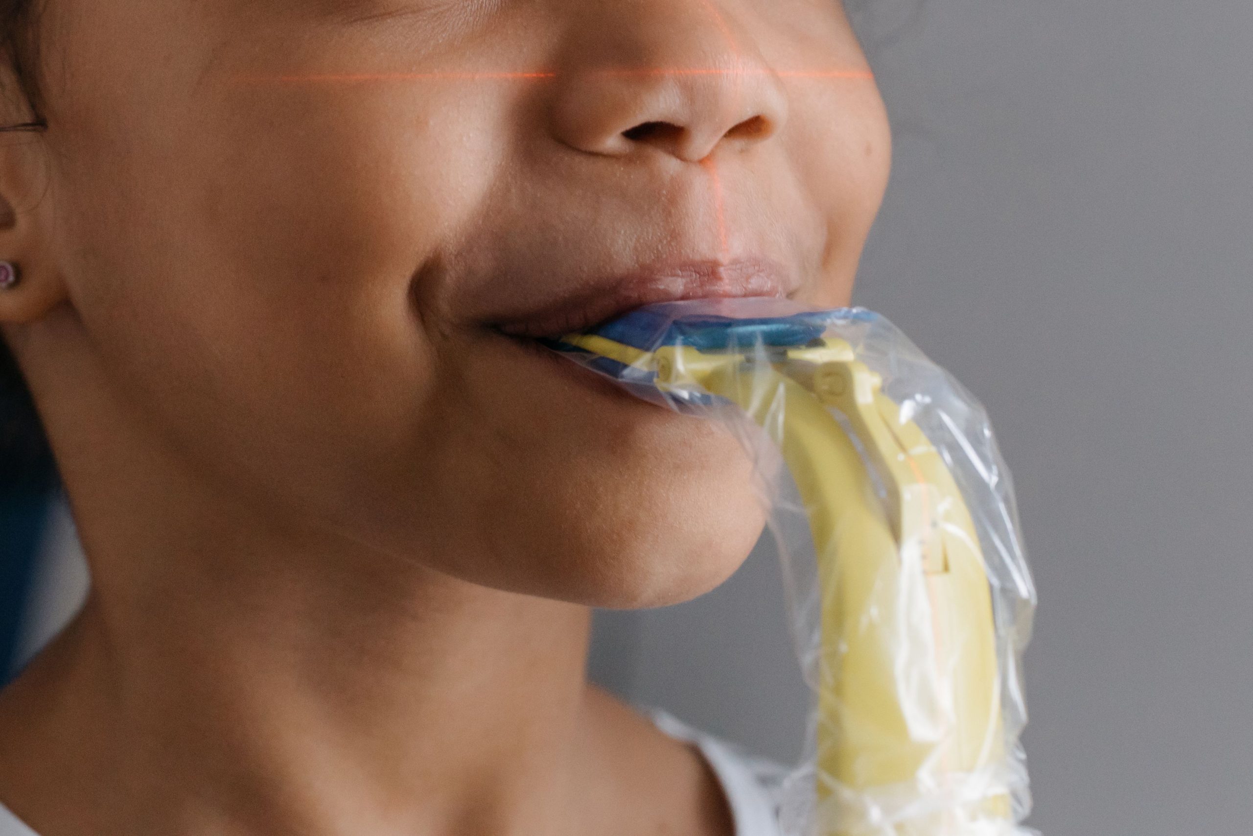
Imagine an 11-year-old child who sleeps with their mouth open every night, wakes up with a dry throat, and struggles to concentrate at school. This is not necessarily just a bad habit. A three-dimensional imaging study published in the Journal of Oral Biology and Craniofacial Research (2025) revealed that mouth breathing in children aged 10–12 years is associated with measurable structural changes: narrower maxillary arches and significantly smaller upper airways compared with children who breathe normally through their noses.
The study was initiated by a team from the Faculty of Dentistry, Universitas Gadjah Mada, led by drg. Anrizandy Narwidina, MDSc, Sp.KGA, Ph.D., as principal investigator and corresponding author from the Department of Pediatric Dentistry. Together with Rani Satiti and drg. Hendri Susanto, the team addressed a question that had remained largely unanswered in Indonesia: precisely how much does chronic mouth breathing affect the craniofacial structures of children?
When the Mouth Takes Over the Nose's Job
Mouth breathing is not a rare phenomenon. In urban Indonesian populations, it is thought to be relatively common, yet high-precision imaging data in the local context have been scarce. This study sought to fill that gap by recruiting 30 children from an elementary school in Kotagede District, Yogyakarta. They were equally divided into 15 children with a mouth-breathing pattern and 15 nasal breathers serving as controls.
The diagnosis of mouth breathing was not based on a single observation. The team used a multi-layered approach involving the Airway Index, Quinn's Nasal Competency Test, Mallampati Classification, tonsillar assessment, the Mirror Test, and a validated questionnaire completed by parents. Following screening, each child underwent Cone Beam Computed Tomography (CBCT), a three-dimensional imaging technology capable of capturing detailed anatomical structures and airway spaces with high accuracy.
The findings were striking. All measured parameters of maxillary arch width, from the molar region to the canine region, were significantly narrower in the mouth-breathing group. The most dramatic narrowing was observed in the intercanine distance: children who breathed through their mouths had an average width of only 21.99 mm, compared with 30.13 mm in the control group. This represented a 27% reduction.
Airway Space That Quietly Disappears
Equally striking findings emerged from measurements of upper-airway volume and cross-sectional area. Oropharyngeal volume in mouth-breathing children was only 24,342 mm³, whereas nasal breathers had a volume of 68,005 mm³. In other words, the airway space in the oropharyngeal region was reduced by as much as 64%.
The cross-sectional area of the nasopharynx was also reduced by almost half: 100.92 mm² compared with 194.06 mm² in the control group, representing a 48% reduction. All of these differences were statistically significant, with p-values below 0.001.
“Mouth breathing in school-aged children is associated with measurable reductions in maxillary arch width and upper pharyngeal airway dimensions.” — Rani Satiti, Hendri Susanto, Anrizandy Narwidina, Journal of Oral Biology and Craniofacial Research, 2025
The researchers explained the mechanism behind these findings. When children breathe through their noses, the tongue rests against the palate and generates lateral pressure that encourages the maxilla to develop outward. When breathing shifts to the mouth, tongue-palate contact is reduced. The outward pressure from the cheeks is no longer balanced, causing the maxillary arch to become narrower. This is known as an imbalance in the orofacial musculature.
CBCT Reveals What Clinical Examination May Miss
One important methodological contribution of this study is its demonstration of the value of CBCT as a diagnostic tool. Three-dimensional reconstruction visually confirmed what the numerical data showed: mouth-breathing children had narrower maxillary arches, while their nasopharyngeal and oropharyngeal air spaces appeared smaller than those of their nasal-breathing counterparts.
Occlusal analysis in the study also found that all children with a nasal-breathing pattern had normal Class I molar relationships, whereas the mouth-breathing group exhibited a range of patterns, including Class II Division 1 malocclusion. This strengthens the argument that mouth breathing is not merely a habit but a functional factor that actively shapes—or more precisely, alters—the development of a child's face and jaws during growth.
The researchers emphasised that ages 10–12 represent a critical window for interceptive intervention. During this stage, craniofacial structures remain highly responsive to functional stimuli. Delayed intervention may have long-term consequences, ranging from permanent malocclusion to sleep disturbances and neurocognitive deficits resulting from intermittent hypoxia.
An Early Warning That Requires Multidisciplinary Action
The study has limitations acknowledged by the researchers themselves: its cross-sectional design does not allow causal inferences, the sample was limited to a single geographical area, and selection bias could not be completely avoided. Future longitudinal and multicentre studies are needed to strengthen these findings.
Nevertheless, the central message does not need to wait for further confirmation. When a child chronically breathes through the mouth, the development of the jaws and airways may not proceed normally. CBCT provides a means of visualising these structural changes before they become permanent.
As emphasised by the research team, management requires collaboration among paediatric dentists, orthodontists, otolaryngologists, and paediatricians. This is not because the problem is procedurally complicated, but because a child's body develops as an integrated whole, and a mouth that remains open every night can affect far more than what is immediately visible.
Source DOI: https://doi.org/10.1016/j.jobcr.2025.07.009
Authors: Nanda Ayu, drg. Achmad Zam Zam Aghasy, M.Kes.
Photo: Pexels