
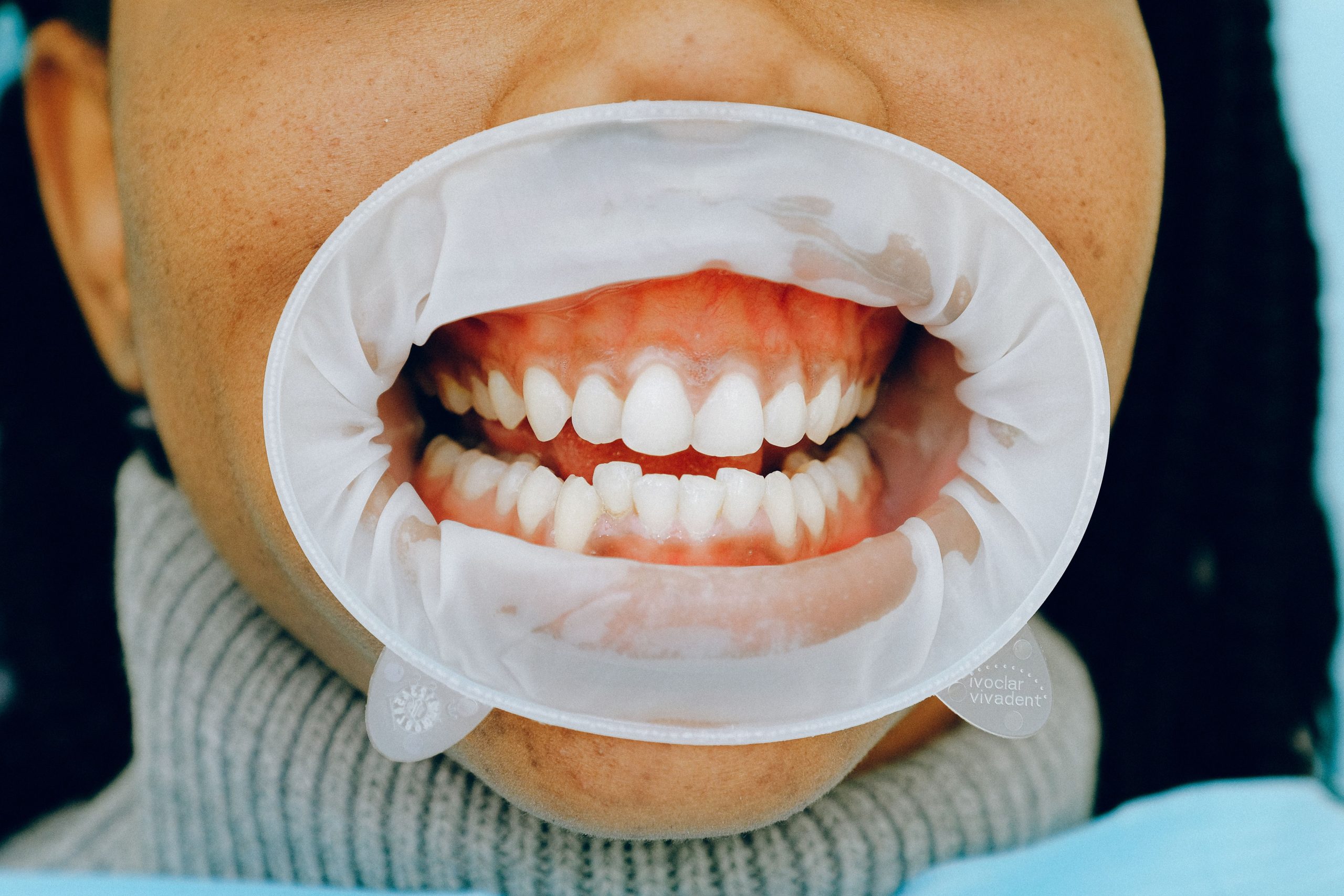
Every time a patient stands in front of a panoramic X-ray machine and rests their chin on the metal support, X-rays rotate around their head for approximately 12 seconds. The dose received is very small, only 47 microsieverts per exposure. But small does not mean zero. In the Dentomaxillofacial Radiology laboratory at the Faculty of Dentistry, UGM, researchers found that even such a small exposure was sufficient to trigger the formation of micronuclei in gingival epithelial cells.
Micronuclei are a marker. They appear when chromosomes within cells are damaged or fail to migrate properly during cell division. The greater the number of micronuclei, the stronger the indication that genotoxic stress is occurring. This raises an important question: can such damage be prevented before exposure occurs?
From the Rabbit Cage to the Dental Chair
Dr. drg. Rurie Ratna Shantiningsih, MDSc, from the Department of Dentomaxillofacial Radiology, Faculty of Dentistry, UGM, had been exploring this question for some time. Her dissertation research used New Zealand rabbits as an initial model, and the results were promising: a mucoadhesive gingival patch containing β-carotene was shown to suppress the increase in micronuclei caused by panoramic radiographic exposure. Moreover, application of the patch did not induce significant inflammatory reactions in the mucosa of the experimental animals.
β-carotene is not a foreign substance. It is a carotenoid pigment abundantly found in carrots and orange-colored vegetables. As an antioxidant, it works by binding free radicals through the double bonds in its structure while also facilitating intercellular communication through gap junctions. This communication is important because gap-junction pathways are weakened or disrupted in many types of cancer cells.
The question then shifted: would what worked in rabbits also work in humans?
Twenty Subjects, One Patch, Ten Days
A clinical study published in the Indonesian Dentistry Magazine in 2015 involved 20 subjects aged 20–25 years who required panoramic radiography for diagnostic purposes. They were randomly divided into two groups: one group received no treatment before exposure, while the other had a β-carotene mucoadhesive gingival patch applied to the anterior maxillary gingival mucosa.
The patch was made from a mixture of HPMC, CMC-Na, propylene glycol, and β-carotene solution. It was designed as a small disc measuring 35 mm in diameter and intended to adhere to the mucosal surface without being easily dislodged by saliva.
Ten days after exposure, gingival mucosal samples were collected from each subject using a cervical brush. The collected cells were then stained using a modified Feulgen-Rossenbeck method and examined under a microscope to determine how many micronuclei had formed.
The results pointed in the right direction. The group using the patch showed a lower mean increase in micronuclei. However, statistically, the difference was not strong enough to be considered significant (p > 0.05).
“Application of a β-carotene mucoadhesive gingival patch to human gingival mucosa has demonstrated a protective role against the effects of panoramic radiographic exposure, although the protection was not yet optimal because statistical analysis showed no significant difference.”
Lasting for More Than Ten Hours, but with a Slightly Uncomfortable Sensation
In addition to micronuclei, the study examined two other aspects: how long the patch could remain in the mouth and whether its presence affected radiographic image quality.
In terms of durability, the results were quite satisfactory. Forty percent of subjects reported that the patch remained attached for more than 10 hours, while 70% were able to retain it for at least more than 5 hours. The backing layer of the patch was designed to prevent water from entering from one side, thereby slowing the swelling process that would normally accelerate the release of the material.
Only one side effect was reported: an uncomfortable foreign-body sensation during the initial period of use. Ninety percent of subjects experienced this sensation, but almost all reported that it gradually disappeared as the mouth adapted to the foreign material on the gingiva. No complaints of heat, itching, or pain were reported.
Equally important, when radiographs from the two groups were analyzed using ImageJ software to compare grayscale values in corresponding areas, no significant differences were found. The patch did not create artifacts, obscure anatomical details, or interfere with clinical interpretation. The alveolar bone, teeth, and surrounding tissues remained clearly visible.
Potential Awaiting Further Refinement
The researchers proposed two possible reasons why the protective effect of the patch did not reach statistical significance. First, the concentration of β-carotene in the formulation was very low, at only approximately 2,290–2,775 micrograms per 50 mm², because β-carotene has very low solubility in alcohol. Second, the patch was applied only once, immediately before exposure, meaning that the antioxidant mechanism may not have had sufficient time to act fully within the mucosal tissue.
The proposed solutions point in two directions: increasing the frequency of application for 10 consecutive days before exposure and developing a nanotechnology-based formulation to improve β-carotene solubility without altering its mucoadhesive properties. Nanometer-scale particles have been shown to enhance the diffusion of active compounds into tissues, as demonstrated in previous toothpaste research.
Ultimately, this study is not about failure. The direction was promising, and the pattern was apparent: a reduction in micronuclei, adequate patch retention, and compatibility with radiographic image quality. What is needed now are higher doses and longer application periods. For patients who require repeated panoramic radiographic examinations, such a small innovation could mean much more than a statistical finding.
Source DOI: https://doi.org/10.22146/majkedgiind.9121
Authors: Anny Anggraini; drg. Achmad Zam Zam Aghasy, M.Kes.
Photo: Pexels