
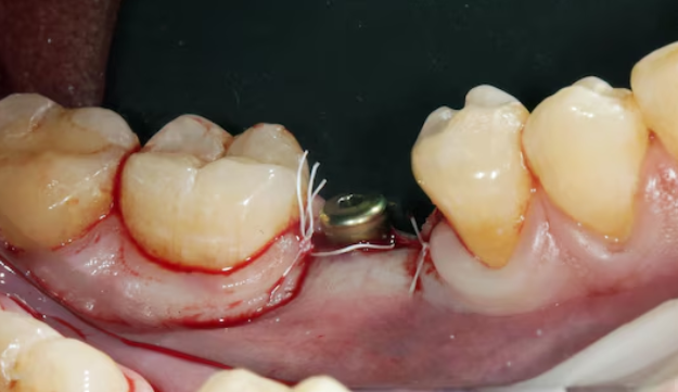
A literature review published in the journal Materials in July 2022 revealed a finding that should change the way we design dental implants: even slight differences in implant surface texture can determine whether harmful bacteria will thrive there or not. The study, led by Dr. drg. Retno Ardhani, M.Sc., from the Department of Dental Biomedical Sciences, Faculty of Dentistry, Universitas Gadjah Mada, examined 26 scientific publications from 2000 to 2021, all focusing on one question: how does Porphyromonas gingivalis, an anaerobic bacterium frequently found at peri-implantitis sites, respond to different surface topographies of subperiosteal implant biomaterials?
Bacteria That Are Smarter Than We Think
Porphyromonas gingivalis is far from an ordinary bacterium. This Gram-negative organism, measuring approximately 1.51 micrometers, possesses several biological weapons: fimbriae, hair-like structures that function as “hooks” for attachment; a protective capsule capable of evading the body's immune system; and aggressive enzymes such as collagenase and hyaluronidase that damage the periodontal tissues surrounding implants.
More concerningly, this bacterium does not only cause localized problems around implants. Research has shown that P. gingivalis can trigger systemic disease through four pathways: bacteremia, or the entry of bacteria into the bloodstream; activation of prolonged inflammatory cascades; toxin dissemination; and direct migration to immune cells throughout the body.
The process of bacterial attachment to an implant surface does not occur instantaneously. A biofilm, a community of bacteria protected by an extracellular polymeric matrix, develops through five stages over a period of one to two weeks and reaches maturity after three months. Once the biofilm has matured, eliminating the bacteria within it becomes far more difficult because they are protected even from antibiotics.
Small Numbers with Major Consequences
Across the 26 studies analyzed, one pattern emerged consistently: the rougher the implant surface, the more P. gingivalis attached to it. The parameter used to measure surface roughness was Ra, the average surface roughness, expressed in micrometers.
On titanium surfaces, studies showed that an Ra roughness above 0.2 micrometers was sufficient to significantly increase the risk of biofilm formation. Rougher surfaces provide more sheltered areas where bacteria can avoid the shear forces of oral fluids while also increasing the contact area between bacteria and the implant.
Conversely, there is an interesting lower threshold: titanium surfaces with an Ra below 0.03 micrometers no longer showed a significant reduction in P. gingivalis attachment. This means that excessively polishing an implant surface does not necessarily provide additional benefits. For titanium, the optimal threshold appears to be around 0.3 micrometers.
“The configuration and size of surface topography affect the attachment of P. gingivalis to subperiosteal implant materials, regardless of the type of material.” — Dr. drg. Retno Ardhani, M.Sc., and research team
Another equally interesting finding comes from studies on nanotopography. Titanium surfaces with 0.5-micrometer-high nanospikes produced using helium ion irradiation were shown to cause physical deformation of P. gingivalis: the bacteria appeared to become “stretched” or “flattened” upon attachment, much like sitting on a bed of tiny spikes. Such bactericidal effects open new possibilities for implant surface designs that actively damage bacteria rather than merely reducing the available sites for bacterial attachment.
It Is Not Just About Roughness
Although surface roughness is a dominant factor, the study emphasized that the picture is not that simple. Two surfaces with nearly identical Ra values may exhibit different bacterial behavior depending on surface chemistry, surface energy, and material hydrophobicity.
A study comparing titanium and zirconia with similar Ra values of approximately 0.21 micrometers found that bovine enamel, which was actually smoother, attracted more P. gingivalis because electrostatic charges on its surface strengthened interactions with bacterial cells. This demonstrates that surface roughness and surface chemistry work together rather than independently.
Another phenomenon was observed in mature biofilms. Once the biofilm had fully developed, the influence of surface roughness decreased, while surface energy became a more important factor in determining the survival of the bacterial colony. The implication is that biofilm prevention strategies must consider the stage of biofilm formation, not merely the initial surface condition.
Zirconia, a material that is increasingly popular as an alternative to titanium, also demonstrated similar behavior. Zirconia-toughened alumina with an Ra of 0.031 micrometers had substantially fewer P. gingivalis bacteria than after sandblasting, which increased the Ra to 0.465 micrometers. Surface treatments such as sandblasting, acid etching, and laser treatment, which have traditionally been used to improve osseointegration—the integration of the implant with bone—also have a direct impact on the risk of bacterial infection.
Designing Smarter Implants
This research carries an important message for clinicians, researchers, and dental implant manufacturers: implant surface design cannot be optimized solely for osseointegration. Every surface modification intended to improve the attachment of bone and soft-tissue cells should also be evaluated for its potential impact on bacterial attachment.
Dr. drg. Retno Ardhani, M.Sc., and the research team recommend that future studies incorporate more comprehensive topographical parameters, such as summit density (Sds) and developed area ratio (Sdr), which can provide a more complete description of the shape and spatial distribution of surface features. These parameters may hold the key to understanding why two surfaces with the same Ra value can behave differently toward bacteria.
A failed dental implant is not merely a financial loss for the patient. Behind every severe case of peri-implantitis lies the risk of progressive bone loss, painful repeat surgical procedures, and even potential systemic effects extending far beyond the oral cavity. Thus, the question of how smooth or rough the surface of a titanium screw measuring only a few centimeters actually is turns out to be anything but a small question.
Authors: Achmad Zam Zam Aghasy, DDS, M.Kes.; Hazra Alifia Muharam
Photo: Freepik
DOI: https://doi.org/10.3390/ma15144988