
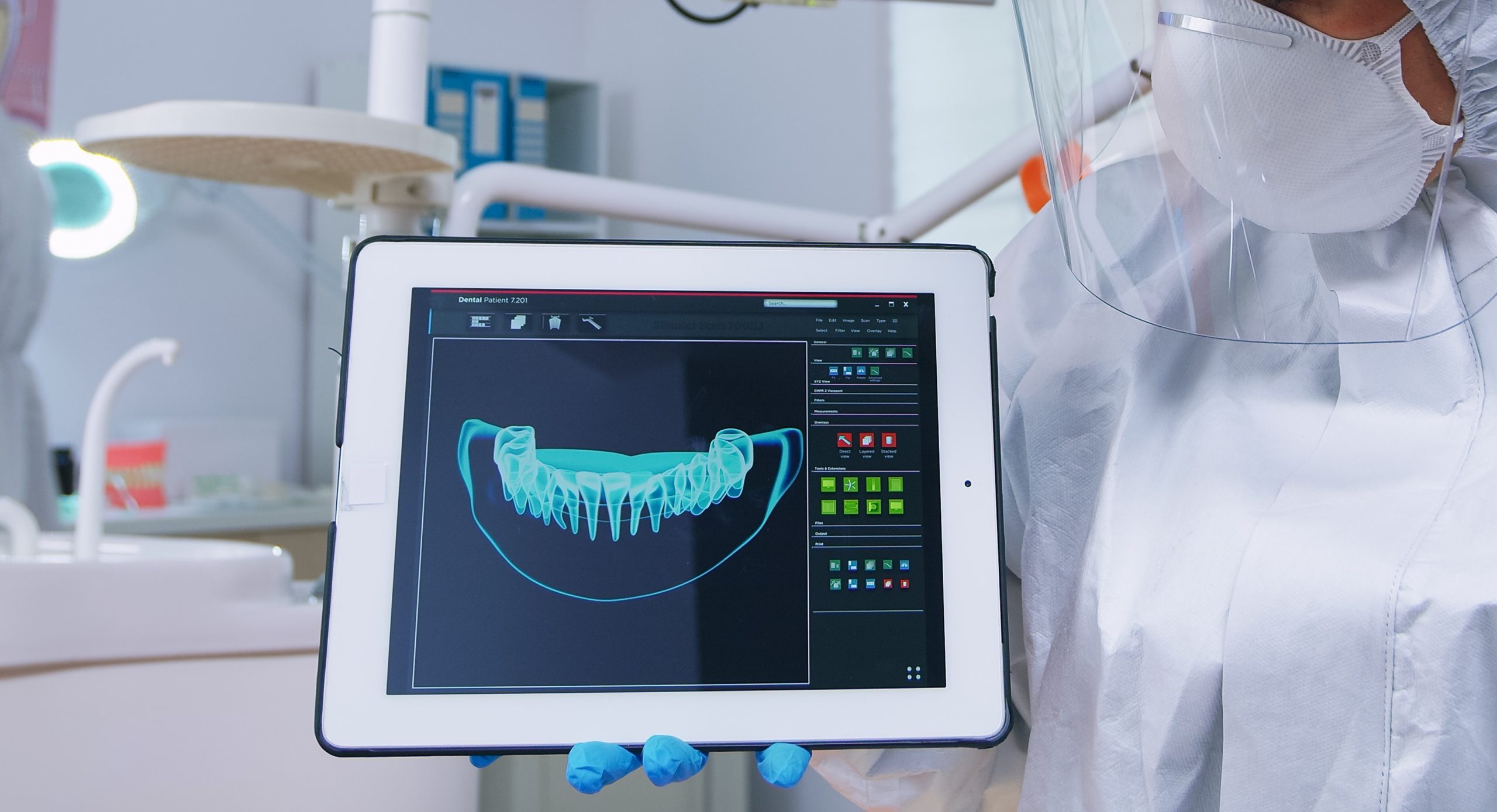
A difference of 1.38 millimeters may seem insignificant. Yet in dentistry, such a small discrepancy can determine whether orthodontic appliances fit correctly, whether dentures are comfortable, or whether a treatment plan proceeds as intended. This seemingly minor measurement difference lies at the heart of a recent study conducted by a multidisciplinary research team from Universitas Gadjah Mada (UGM), which systematically compared three methods of measuring dental study models.
Published in the International Journal of Community Dentistry in 2025, the study involved researchers from UGM's Department of Mechanical and Industrial Engineering, Department of Pediatric Dentistry, Department of Oral and Maxillofacial Surgery, Department of Biomedical Sciences, and Department of Prosthodontics. One of the contributors from Pediatric Dentistry was Dr. drg. Indra Bramanti, M.Sc., Sp.KGA(K), who collaborated with eight other researchers to investigate a fundamental question: How reliable are digital measurement methods compared with conventional techniques?
Three Measurement Methods, One Reference Standard
The researchers evaluated 15 dental study models, consisting of seven mandibular and eight maxillary models, using three different measurement techniques.
The first method served as the reference standard. A transparent acetate sheet was placed over each model, anatomical landmarks were marked, and measurements were taken using a precision Vernier caliper. The second method used digital photography. A digital camera was positioned 5 centimeters above the occlusal surface of each model, with a ruler included for calibration. Measurements were then performed using image analysis software. The third method employed the Aoralscan 3 Wireless Intraoral Scanner (Shining 3D), which generated three-dimensional digital models for further analysis. Aoralscan 3 Wireless Intraoral Scanner buatan Shining 3D, yang menghasilkan model 3D terdigitalisasi untuk dianalisis lebih lanjut.
The findings were clear. Compared with manual measurements, the digital scanner produced a mean absolute error (MAE) of only 0.91 mm, whereas the photographic method recorded an MAE of 2.29 mm. In terms ofmean relative error(MRE), digital scanning achieved an error rate of 13%, compared with 31% for photographic measurements.
Mandibular Models Pose Greater Challenges
Not all dental arches proved equally easy to scan. One notable finding was the difference in scanning accuracy between mandibular and maxillary models. Digital scanning produced greater deviations in mandibular models, likely due to their more complex anatomical contours. For the best-performing maxillary model, the scanner achieved an MAE of only 0.40 mm with an MRE of 5%. In contrast, the most challenging mandibular model recorded an MAE of 1.85 mm and an MRE of 26%.
The researchers suggested that the more intricate surface topography of the mandible contributes to increased measurement variability.
The photographic method, meanwhile, demonstrated consistent limitations across both mandibular and maxillary models. Image resolution and dependence on camera positioning remained major sources of measurement error. Even under optimal conditions, the photographic method never achieved an MAE below 1.97 mm.
"Intraoral scanning represents a more robust and dependable modality for precise dental model assessment." — International Journal of Community Dentistry, 2025
From Plaster Casts to Digital Dentistry
For decades, plaster study models have been the foundation of orthodontic diagnosis and treatment planning. They are inexpensive, durable, and capable of capturing fine anatomical detail. However, producing and measuring them requires considerable time and labor. Digital intraoral scanning offers several advantages, including improved patient comfort, shorter clinical procedures, and the elimination of distortions associated with conventional impression materials.
Beyond demonstrating superior statistical accuracy, the study also showed that intraoral scanners provide better repeatability , as reflected by consistently lower standard deviations than the photographic method. In other words, digital scanners not only produce more accurate measurements but also deliver more consistent results across repeated assessments.
The research was supported by the Kedaireka Matching Fund Program of the Directorate General of Higher Education, Research, and Technology, Ministry of Education, Science, and Technology of the Republic of Indonesia.
For clinicians who continue to rely on occlusal photographs for quantitative measurements, these findings offer an important reminder. Photography may be simple and inexpensive, but a 31% relative measurement error can be a costly compromise in clinical practice.
Source DOI: https://doi.org/10.56501/intjcommunitydent.v13i1.1280
Authors: Nanda Ayu, drg. Achmad Zam Zam Aghasy, M.Kes.
Photo: Freepik