
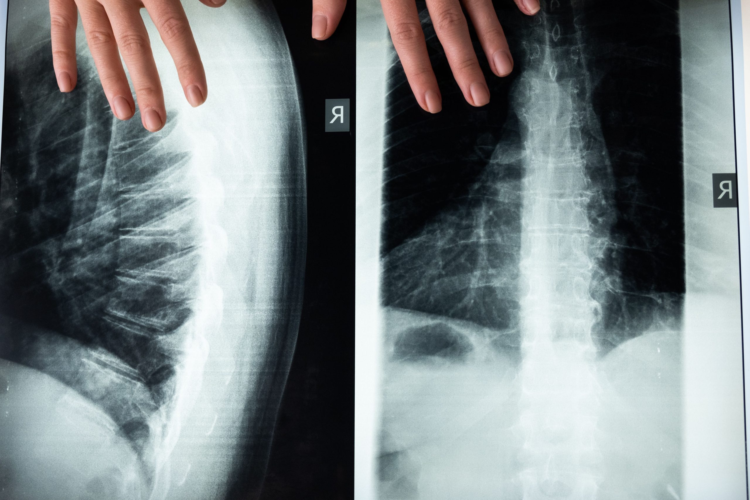
Imagine a pregnant mother whose protein intake is far below adequate levels. Her baby is born, grows, and eventually begins receiving nutritious food. Can the child's bones “catch up” with the earlier deficiency? Research by Prof. Dr. drg. Pinandi Sri Pudyani, SU., Sp.Ort(K), of the Department of Orthodontics, Faculty of Dentistry, Universitas Gadjah Mada, provides a surprising answer: no.
Impaired bone calcification caused by protein deficiency from the prenatal period through breastfeeding was found to be irreversible. Providing adequate protein after weaning was unable to restore bone development that had already been compromised.
When the Bone Matrix Fails to Develop
Bone is not merely a passive framework. It is living tissue that is continuously formed, remodelled, and mineralised. Bone calcification—the process by which calcium and phosphorus minerals are deposited into the organic bone matrix—is an indicator of skeletal maturity. In orthodontics, skeletal maturity is not merely supplementary information; it determines when and how treatment should be performed, from the use of functional appliances to orthognathic surgical planning.
Protein plays a dual role in this process. First, it serves as the building material for the organic bone matrix, which is composed primarily of type I collagen. Second, approximately 40% of plasma calcium is bound to proteins as a reserve. Therefore, inadequate protein intake can impair mineral deposition into bone through two pathways simultaneously.
Pudyani designed a study involving 30 Rattus norvegicus rats divided into three groups. The first was the control group, in which both the mothers and offspring received a standard diet containing 25% protein. The second group experienced severe protein deficiency, from pregnancy through adulthood at 56 days of age, receiving a diet containing only 4–10% protein. The third was the “recovery” group: the mothers experienced protein deficiency, but their offspring were given a standard diet from weaning at 30 days of age until adulthood.
Two parameters were measured. Histologically, the width of the right femoral epiphyseal plate was measured in micrometres following haematoxylin-eosin staining. Biochemically, calcium and phosphorus levels in the left femur were measured using atomic absorption spectroscopy and ultraviolet-visible spectrophotometry.
The Numbers Speak Loudly
The results left little room for ambiguous interpretation.
In the control group, the mean epiphyseal plate width was only 143.40 micrometres, indicating that the bones were approaching maturity, with normal epiphyseal-diaphyseal fusion. The protein-deficient group (Group II) had an epiphyseal width of 294.00 micrometres—almost twice as large—indicating bones that were far from mature. Surprisingly, the recovery group (Group III) recorded an even larger value of 332.5 micrometres and was not significantly different from Group II (p > 0.05).
Bone mineral levels reinforced the same pattern. Calcium levels in the control group reached 30.357 μg/100 g of sample weight. The protein-deficient group had only 10.715, while the recovery group, despite receiving a nutritionally adequate diet for 26 days, reached only 14.40. Bone phosphorus followed a similar pattern: 10.540 in the control group, 3.861 in the protein-deficient group, and 5.908 in the recovery group.
“Providing a standard diet from weaning through adulthood cannot correct the impairment of bone calcification caused by pre- and postnatal protein deficiency; therefore, the impairment is irreversible.” — Prof. Dr. drg. Pinandi Sri Pudyani, SU., Sp.Ort(K)
One-way ANOVA and t-tests confirmed significant differences (p < 0.01) between the control group and both treatment groups across all measured parameters.
A Window That Closes Before It Can Be Reopened
Why did recovery fail to occur? The answer lies in the biology of cellular development. During early growth, organs undergo a phase of hyperplasia, characterised by rapid and extensive cell division. If this critical period is disrupted in a way that inhibits DNA replication, tissues lose their capacity to increase their number of cells. This damage cannot subsequently be compensated for because the biological “window” has already closed.
Protein deficiency inhibits cellular differentiation and the synthesis of the organic bone matrix. As a result, minerals have no “home” in which to be deposited. Even when adequate protein becomes available later, the foundation that should have been established during the prenatal period is no longer present.
Histological findings showed that Groups II and III had both failed to form an epiphyseal line, indicating that fusion between the epiphysis and diaphysis of the femur had not occurred. Both groups also showed abundant trabecular bone with reduced cortical bone, a pattern reflecting poor bone quality.
Implications Beyond the Laboratory
For orthodontists, these findings have clear clinical relevance. Skeletal maturity determines the optimal timing of treatment, including the correction of mandibular growth discrepancies and the maintenance of occlusal stability after treatment. If bone calcification has been disrupted from an early stage, assessments of skeletal maturity using cervical vertebral analysis or hand radiography may provide misleading information if the patient's nutritional history is not taken into consideration.
More broadly, this study reminds us that a child's dental and craniofacial health may be determined long before the child sits in an orthodontic chair—even before birth. Maternal nutrition is not merely a matter concerning pregnancy; it is an investment in the skeletal architecture that will support a child's face, teeth, and smile throughout life.
Pudyani concluded her research by recommending further studies on other nutrients, including magnesium, zinc, and iodine, as well as their effects on facial soft tissues. More questions remain than have been answered. But one thing is certain: some damage does not wait to be treated because it may already be fully established before we even know it exists.
Authors: Nanda Ayu, drg. Achmad Zam Zam Aghasy, M.Kes.
Photo: Pexels