
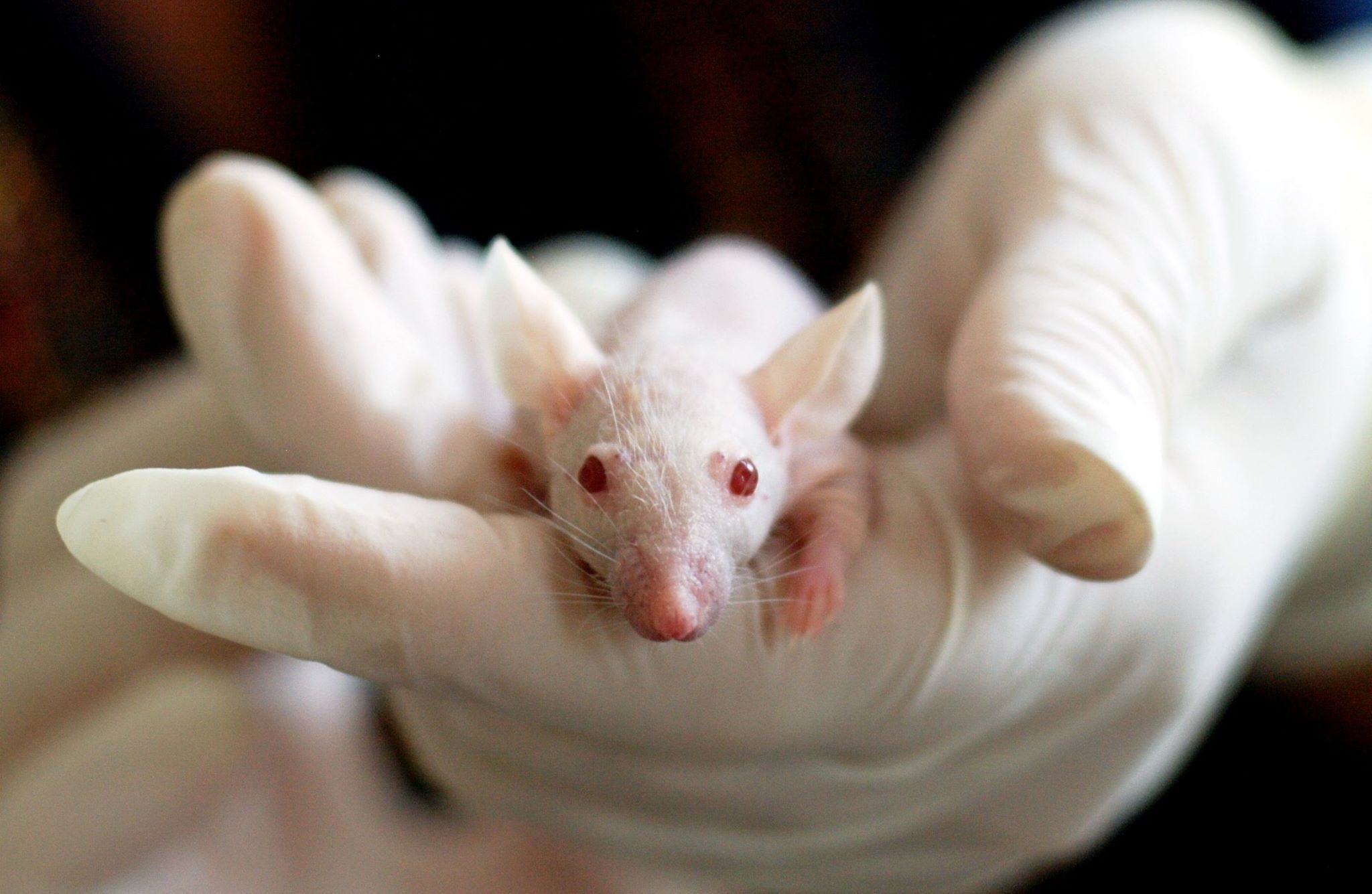
Of 54 experimental mice carrying the v-H-ras oncogene, 47 developed spontaneous tumors—not in the areas being investigated, but in unexpected locations: swollen jaws, fractured lower incisors, and papillomas growing around the mouth and on the back. These findings emerged from research by Prof. Dr. drg. Dewi Agustina, M.D.Sc., a lecturer in the Department of Oral Medicine, Faculty of Dentistry, Universitas Gadjah Mada, published in the Indonesian Journal of Dentistry in 2009.
The study was not merely about what the researchers had planned to investigate. Rather, the unexpected findings were what ultimately told the most compelling story.
When an Oncogene Speaks Louder Than Expected
The conceptual starting point of the study was straightforward: would transgenic mice carrying the v-H-ras gene be more susceptible to developing oral mucosal carcinoma when exposed to promoting agents such as 4-nitroquinoline 1-oxide (4NQO) and phorbol 12,13-didecanoate (PDD) applied to the palate?
The H-ras gene was selected for a reason. Mutations at codons 12 and 61 of this gene had previously been detected in the oral squamous epithelium of mice and rats induced with 4NQO, making it a strong candidate for an initiating gene in oral carcinogenesis. The logic was straightforward: if cells had already been “initiated” by the transgene, exposure to a promoting agent should accelerate the progression toward dysplasia and carcinoma.
The results were surprising. After 24 weeks of 4NQO administration, there was little evidence of palatal epithelial dysplasia, and the changes that did occur were mild. Only 6 of 106 mice treated with 4NQO exhibited dysplastic changes, and only two developed moderate dysplasia. Neither the PDD nor PD groups showed any dysplasia.
“Activated v-H-ras did not increase the rate of palatal mucosal carcinogenesis in the model used.”
In other words, the primary hypothesis was not confirmed. Yet this is precisely where science reveals its true nature.
Tumors That Arrived Uninvited
What drew attention instead were the incidental abnormalities that developed outside the area under investigation. Of the 54 transgenic mice, 47 developed one or more spontaneous abnormalities: cutaneous papillomas around the mouth, on the back, and in the anal region; visibly enlarged mandibles; lower incisor fractures first detected when the mice were 13 to 17 weeks old; and soft-tissue growths in various locations, including the parotid glands, thoracic cavity, and floor of the mouth.
Histopathological examination revealed a diverse range of lesions, including squamous cell carcinoma, lymphoma, and malignant mesenchymoma, as well as one case of squamous cell carcinoma with an inverted papillary configuration in the floor of the mouth. Radiographic examination of the enlarged mandibles revealed bone expansion with a multilocular pattern, resembling the appearance of an aggressive odontogenic tumor.
One particularly novel finding was the recurring pattern of lower incisor fractures in the transgenic mice, while none of the control mice experienced such fractures. Histologically, these teeth exhibited features resembling Type I dentinal dysplasia in humans, an autosomal dominant condition whose etiology remains incompletely understood. Prof. Dewi noted that this finding could potentially provide an animal model for studying this developmental disorder of dentin.
By comparison, only four of the 54 control mice—the parental FVB/N strain without the transgene—developed incidental abnormalities, all of which were mild.
Why This Matters for Oral Cancer Research
The research was conducted at the School of Dental Science, University of Melbourne, and the Ludwig Institute for Cancer Research in Victoria, Australia, with funding support from AIDAB. Ethical approval was obtained from two animal ethics committees in Australia. Following euthanasia, all tissues were processed using standard H&E staining, while palatal epithelial dysplasia was assessed using a modified semiquantitative method developed by Smith and Pindborg.
Based on the overall findings, Prof. Dewi drew an important conclusion: the differences between this study and previous studies using similar transgenic mice may have resulted from differences in the site of transgene insertion within the chromosome. Different insertion sites of the same transgene can produce dramatically different patterns of abnormalities because oncogene expression is strongly influenced by the genomic context surrounding the insertion site.
More importantly, the emergence of numerous spontaneous abnormalities in transgenic mice carrying a single oncogene raised another unanswered question: could other oncogenes have been activated in addition to the v-H-ras transgene itself? Prof. Dewi noted that this possibility would need to be confirmed through oncogene analysis of the tumor tissues.
This study is therefore more than a negative report of an unconfirmed hypothesis. It is a reminder that in cancer biology, “incidental findings” often contain deeper questions than those researchers initially bring into the laboratory.
DOI: —
Authors: Anny Anggraini; drg. Achmad Zam Zam Aghasy, M.Kes.
Photo: Pexels