
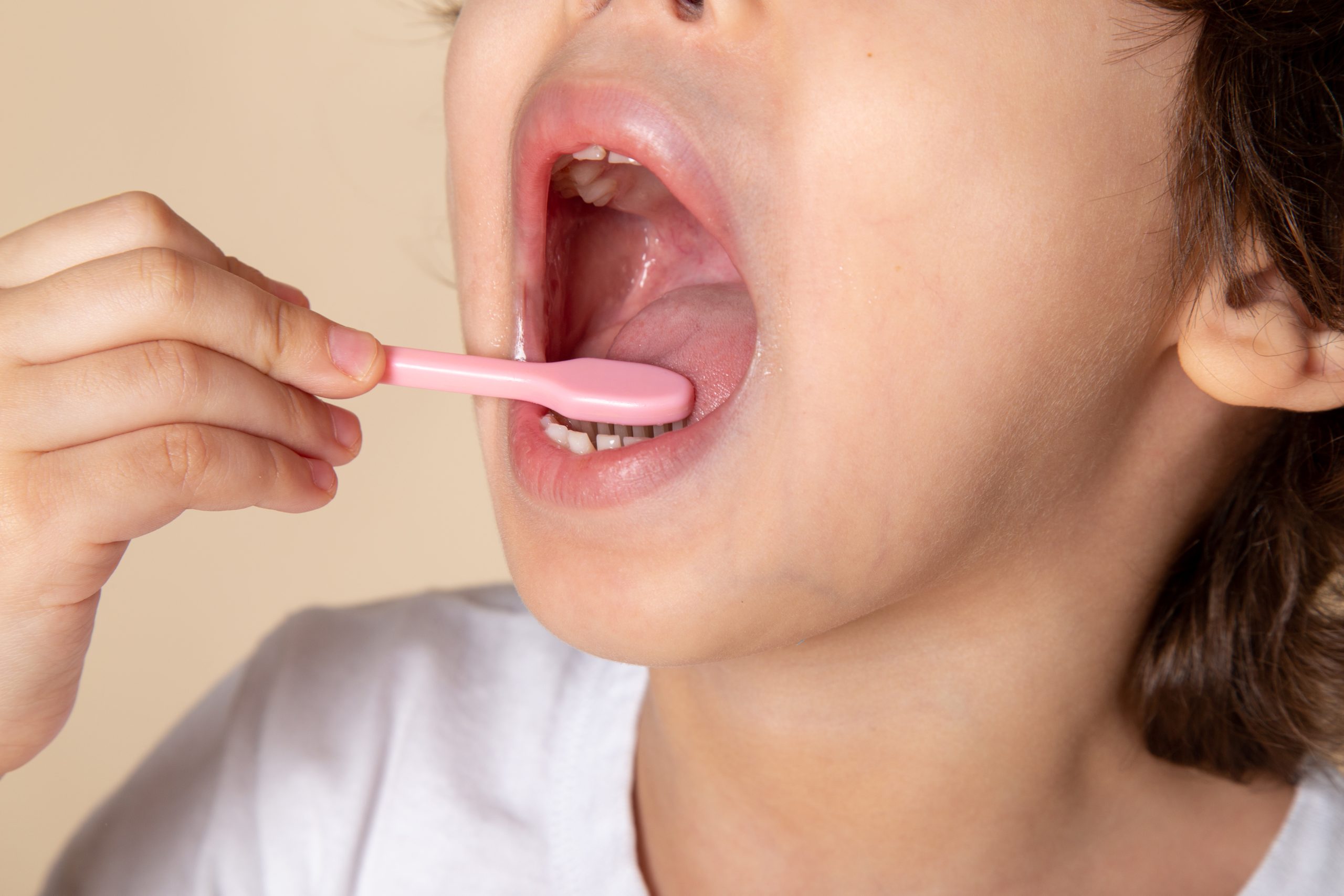
The figure is striking: up to 95 percent of the population has experienced traumatic ulcers, wounds of the oral mucosa commonly referred to as canker sores. Most people consider them minor, wait for them to heal on their own within two weeks, or apply an over-the-counter gel from a pharmacy. Yet behind this seemingly simple condition lies a complex biochemical process of oral mucosal wound healing involving four sequential phases that must occur at precisely the right time.
Prof. Dr. drg. Juni Handajani, M.Kes., Ph.D., PBO, from the Department of Oral Biology, Faculty of Dentistry, Universitas Gadjah Mada, asked a more fundamental question: what if this process could be accelerated—not with synthetic chemical drugs, but with biological signals derived from stem cells found in children’s baby teeth?
The Secret Inside a Lost Baby Tooth
Baby teeth shed by children around the age of six turn out to contain something valuable. Within their dental pulp live a population of stem cells known as Stem Cells from Human Exfoliated Deciduous Teeth, or SHED. These cells are more than ordinary “reserve cells.” SHED can differentiate into fibroblasts, odontoblasts, adipocytes, nerve cells, and epithelial cells, while secreting various important growth factors such as FGF, TGF, CTGF, NGF, and BMP.
What is particularly interesting is not the cells themselves, but what they release. When the stem cells are cultured in serum-free medium, the resulting spent culture medium, known as SHED-SM, contains a rich mixture of growth factors, cytokines, chemokines, extracellular vesicles including exosomes, and other bioactive molecules. This collection of secreted factors is known as the secretome, and it is the focus of this research.
Using intact stem cells as a therapy carries its own risks, including the possibility of cellular mutations leading to tumorigenesis and the risk of rejection following transplantation. Spent medium offers a middle ground: the biological benefits of the secretome without the risks associated with implanting living cells.
From Dental Pulp to Nanoemulgel
The research team, led by Prof. Dr. drg. Juni Handajani, M.Kes., Ph.D., PBO, together with collaborators from Universitas Airlangga and Universitas Indonesia, did not stop at isolating the secretome. They formulated SHED-SM into a nanoemulgel, a topical preparation that combines the advantages of nanoemulsions and gels: nanoparticles for optimal absorption and a gel consistency that makes application to wounded mucosal surfaces easier.
The study was funded through the Indonesian Collaborative Research Program under contract number 1917/UN1/DITLIT/PT.01.03/2024 and received ethical approval from the Ethics Committee of FKG-RSGM Prof. Soedomo UGM. Before being tested in animals, the SHED-SM nanoemulgel was evaluated for its effects on the viability of NIH-3T3 fibroblast cells using an MTT assay.
The results showed that SHED-SM H1, the medium collected on the first day of incubation, produced the highest cell viability at 65.95 percent, substantially higher than SHED-SM H5, which reached only 37.01 percent. The difference was statistically significant and provided the basis for selecting the formulation for the in vivo stage.
Rats, Three-Millimeter Wounds, and the Critical Fifth Day
Thirty male Wistar rats aged two to three months were divided into two groups: a treatment group receiving SHED-SM nanoemulgel and a control group receiving Aloclair Plus. Standardized 3-mm-diameter traumatic ulcers were created using a punch biopsy on the right buccal mucosa, after which the gel was applied once daily for four consecutive days.
Observations were conducted on days 1, 3, 5, 7, and 14, including clinical evaluation, histological examination using Hematoxylin-Eosin staining, and immunohistochemistry to measure the expression of COX-2, Collagen-1 (Col-1), and VEGF.
The results spoke for themselves. By day five, the epithelium in the treatment group had already closed, while the control group had not. By day seven, the epithelium in the treatment group appeared thicker than that of the control group.
“Application of SHED Spent Medium nanoemulgel can accelerate the healing of traumatic buccal mucosal ulcers, as demonstrated by epithelial closure and increased neutrophil counts and expression of COX-2, Col-1, and VEGF.” — Prof. Dr. drg. Juni Handajani, M.Kes., Ph.D., PBO, in the study’s conclusion published in F1000Research, 2025
At the molecular level, the mechanism is multifaceted. SHED-SM recruits neutrophils and monocytes during the early inflammatory phase and subsequently promotes the polarization of macrophages from the pro-inflammatory M1 phenotype toward the reparative M2 phenotype. Growth factors such as VEGF and FGF in SHED-SM stimulate endothelial-cell proliferation to form new blood vessels, while TGF-β modulates collagen synthesis and tissue remodeling.
When Canker Sores May No Longer Be So Trivial
The 95 percent prevalence of traumatic ulcers reported in the 2018 Riskesdas survey is more than just a statistic. It means that almost everyone experiences the condition at some point, with many people experiencing recurrent episodes. Most treatments currently available are symptomatic: they reduce pain, prevent infection, and allow time to take its course.
This research opens up another possibility. Baby teeth that are typically discarded or kept as childhood keepsakes could potentially become a source of regenerative therapy that is safe, practical, and affordable. Spent medium does not require cell transplantation, does not carry the same tumorigenesis risk associated with implanting living cells, and can be obtained through a relatively simple process.
There is, of course, still a significant gap between the laboratory and clinical application. Animal testing is an initial step, not the final one. Nevertheless, the direction indicated by this research gives us reason to think twice before simply throwing away a baby tooth that has just fallen out.
Source DOI: https://doi.org/10.12688/f1000research.164339.1
Authors: Anny Anggraini; drg. Achmad Zam Zam Aghasy, M.Kes.
Photo: Freepik