
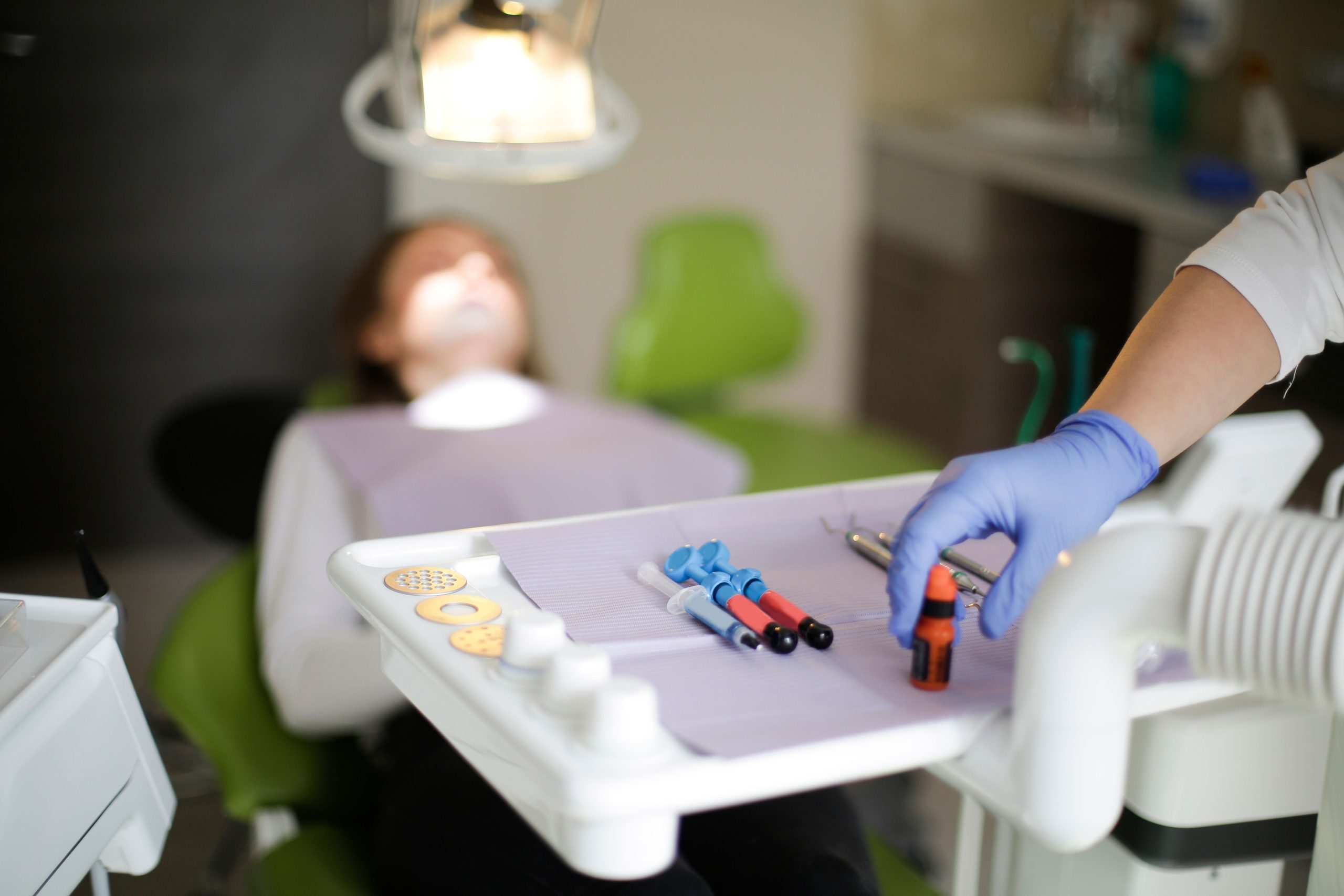
Two days after falling while running in her home yard, a four-year-old girl arrived at Prof. Soedomo Dental Hospital, Faculty of Dentistry, Universitas Gadjah Mada. She was crying. Her upper front tooth was broken and bled whenever it was touched, and she refused to eat because she was afraid of the pain. Her mother had already given her pain medication, but it provided little relief.
The case subsequently became the subject of a scientific case report published in e-GiGi (Volume 12, Issue 1, 2024) by drg. Suci N. Rahmadani and drg. Putri Kusuma Wardani Mahendra, M.Kes., Sp.KGA., from the Department of Pediatric Dentistry at FKG UGM. Their findings demonstrated that a complex crown fracture in a primary tooth—even in an uncooperative child—can be successfully managed with good outcomes when the appropriate approach is used and collaboration among the dentist, parents, and child is strong.
When a Baby Tooth Cannot Simply Be Left Untreated
A complex crown fracture occurs when a tooth fracture extends into the pulp, the innermost part of the tooth containing nerves and blood vessels. This is far more serious than an ordinary cracked tooth.
According to data cited in the case report, dental trauma in children aged 2 to 4 years occurs in approximately 20% of cases, with the maxillary central incisors being the most frequently affected teeth. Their prominent position in the oral cavity makes them particularly vulnerable when children fall. In this case, teeth 51 and 61, the left and right maxillary central incisors, respectively, sustained crown fractures with exposed pulp.
What makes the condition urgent is not only the pain. Once exposed, the pulp becomes an entry point for bacterial infection. Untreated infection can damage the developing permanent tooth underneath, cause malocclusion as a result of premature tooth loss, and potentially affect speech and the child’s psychological well-being.
The More Ambitious Option: Vital Pulpectomy
Faced with this condition, drg. Putri Kusuma Wardani Mahendra, M.Kes., Sp.KGA., and her team had several options: pulpotomy, vital pulpectomy, or extraction. Pulpotomy, which removes only the coronal portion of the pulp, is appropriate when inflammation remains reversible. In this case, however, the fracture had occurred two days earlier and was accompanied by a history of spontaneous pain and persistent bleeding—signs suggestive of irreversible inflammation.
The team therefore chose vital pulpectomy, which involves removing the entire coronal and radicular pulp tissue, followed by cleaning and filling the root canals. One study cited in the report found a success rate of 96% for vital pulpectomy, compared with 75% for pulpotomy in cases of complex crown fractures.
“The benefit of performing vital pulpectomy on primary teeth with crown fractures and exposed pulp is to prevent malocclusion caused by premature tooth loss, maintain masticatory function, prevent speech problems, prevent psychological effects, and prevent disturbances in the eruption of permanent teeth.” — Rahmadani & Mahendra, e-GiGi, 2024
The procedure was not simple. The first visit began with sterilization and topical anesthesia, followed by infiltration and intrapulpal anesthesia. The pulp tissue was removed using a barbed broach, and the root canals were irrigated with 2.5% NaOCl and saline. Calcium hydroxide paste was then placed as an intracanal dressing. During the second visit, the root canals were obturated using a combination of calcium hydroxide and iodoform paste (Metapex), selected for its biocompatibility, resorbability, and lack of toxicity to periapical tissues and the developing permanent tooth.
A Crying Child and a Mother’s Embrace: Behavior Management as the Key
The greatest challenge in this case was not the clinical technique but the psychological aspect. The child was classified as “definitely negative” according to the Frankl behavior scale, cried hysterically, and refused all treatment. A radiograph could not even be obtained during the first visit because of her condition.
The team chose a non-pharmacological body-restraint approach: the child’s mother sat in the dental chair while the child lay on her lap, with the mother securely holding the child’s body, arms, and legs throughout the procedure. This technique is not simply a matter of “forcing” the child to remain still; it is a structured clinical approach used to prevent injury and allow treatment to be performed safely.
The approach also reflected a principle emphasized in the report: successful management of dental trauma in children depends not only on the dentist’s clinical skills but also on active parental involvement, from the initial explanation and informed consent through to the actual procedure.
The result? By the third visit, the child had no complaints. Clinical examination showed negative findings for percussion, palpation, and mobility. The final restoration was successfully placed using Type II Glass Ionomer Cement (GIC). One month later, the child was able to bite food comfortably using her front teeth.
Source DOI: https://doi.org/10.35790/eg.v12i1.47582
Authors: Nanda Ayu, drg. Achmad Zam Zam Aghasy, M.Kes.
Photo: Pexels