
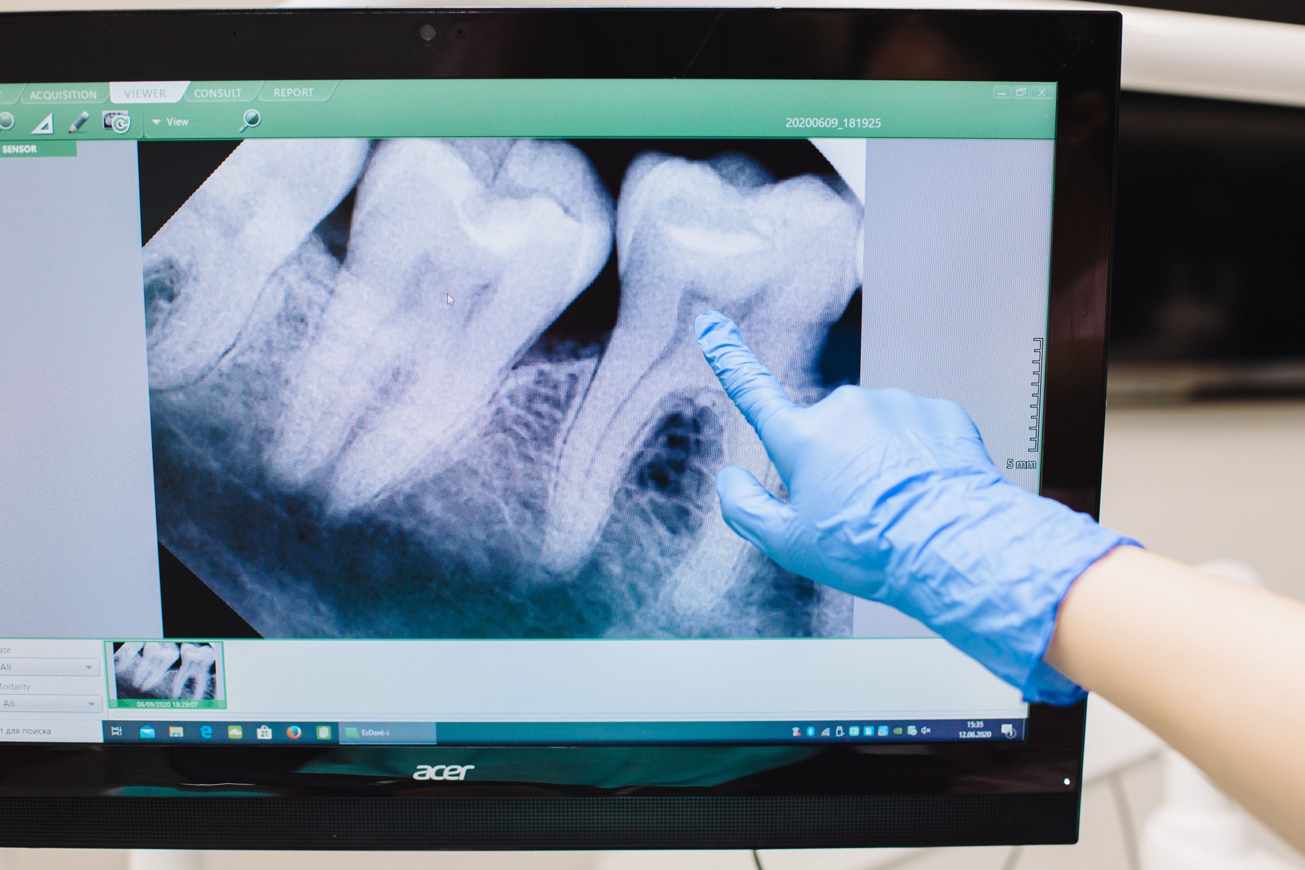
Imagine a 60-year-old woman visiting a dental clinic for a routine dental examination. The dentist takes a periapical X-ray, a procedure that is quick, inexpensive, and routinely performed. Yet, beyond revealing the condition of the tooth roots, that black-and-white image may contain much more information: clues about overall bone density, including a potential risk of osteoporosis that has not yet been detected.
This is the essence of a study published in the Journal of Oral Biology and Craniofacial Research in 2025, involving Dr. drg. Rurie Ratna Shantiningsih, MDSc, from the Department of Dentomaxillofacial Radiology, Faculty of Dentistry, Universitas Gadjah Mada, together with a research team from Universitas Airlangga. The study explored the potential use of trabecular alveolar bone pattern analysis on periapical radiographs as an early marker of osteoporosis, particularly in postmenopausal women.
Why the Jawbone Can Reflect the Condition of Bones Throughout the Body
Osteoporosis is a condition characterized by reduced bone density, making bones fragile and more susceptible to fractures. More than 200 million women worldwide are estimated to be affected by this condition, with the risk increasing sharply after menopause as estrogen levels decline substantially. The gold standard for diagnosis is Dual-Energy X-ray Absorptiometry (DXA), which measures Bone Mineral Density (BMD). However, DXA is expensive, not always readily available, and does not provide detailed information about bone architecture.
This is where dental radiography presents an intriguing alternative. Periapical X-rays, which are commonly performed in dental clinics, can visualize the trabecular structure of the mandibular alveolar bone. Using Fractal Dimension (FD) analysis, the complex pattern of this trabecular network can be measured quantitatively.
The research team analyzed 93 periapical radiographs from 31 postmenopausal women aged over 55 years. Each participant also had BMD data obtained through DXA examination. The radiographic data were obtained from Prof. Soedomo Dental Hospital, UGM, while the BMD data were obtained from patient medical records at Dr. Sardjito General Hospital, Yogyakarta.
One Small Number, One Major Clue
The study measured two main parameters through FD analysis: Alveolar Bone Density (ABD), defined as the ratio of bone area to the total area analyzed, and D value, defined as the number of terminal trabecular points per square centimeter. Both parameters were measured in the anterior region (mandibular symphysis) and posterior region (distal to the mental foramen) using ImageJ software.
The findings were notable in two respects. First, no significant difference was found between FD values in the anterior and posterior regions. This suggests that osteoporosis-related reductions in bone density among postmenopausal women occur relatively uniformly throughout the jaw. This finding is consistent with the theory that estrogen deficiency has systemic effects, influencing the distribution of blood flow and nutrients to bone in a relatively uniform manner.
Second, and more importantly from a clinical perspective, ABD showed a significant positive correlation with BMD status (r = 0.383, p < 0.05). The higher the ABD value, the better the overall bone mineral density. More specifically, ABD in the anterior mandibular region was significantly correlated with the T-score (r = −0.357, p < 0.05), a score used to diagnose osteoporosis according to World Health Organization (WHO) standards.
“ABD may serve as a potential indicator for detecting the risk of osteoporosis in postmenopausal women, particularly in the anterior region, using periapical radiographs.”
Meanwhile, D value showed no significant correlation with either BMD or T-score. The researchers suggested that this may be related to the inherent limitations of two-dimensional radiographs: overlapping trabeculae make it difficult to accurately count terminal points, making D value less reliable in this context.
From the Dental Chair to Early Osteoporosis Detection
These findings have potentially broad practical implications. Most participants in the study had characteristics that reflect the conditions of older women in Indonesia: 71% were physically inactive, 71% had never consumed milk, 90.3% had never received calcium therapy, and 45.1% were classified as severely obese based on BMI. Nevertheless, most participants (61.29%) were classified as having osteopenia based on DXA, while 29.03% had already reached the osteoporosis category.
These figures illustrate the substantial burden of undiagnosed osteoporosis in the community and highlight the strategic position of dental clinics as potential points of first contact with the healthcare system. Periapical X-rays are already routinely performed; the only additional step required would be FD analysis using freely available software.
The researchers also acknowledged several limitations. The small sample size (31 participants) reduced the statistical power of the study. No longitudinal data were available, meaning that changes in bone density over time could not be evaluated. Variations in intraoral conditions among participants were also not fully controlled. Larger studies with prospective cohort designs will be needed before this method can be recommended as a standard screening protocol.
Nevertheless, the direction is clear. X-rays, which have traditionally been viewed primarily as dental diagnostic tools, are gradually taking on a new role: serving as a small window that may provide clues about the bone health of aging women long before a fracture occurs.
Source DOI: https://doi.org/10.1016/j.jobcr.2025.06.025
Authors: Anny Anggraini; drg. Achmad Zam Zam Aghasy, M.Kes.
Photo: Pexels