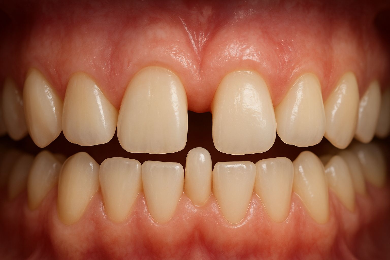
Supernumerary teeth are additional teeth that appear in excess of the normal number in the permanent dentition. This condition is often associated with congenital disorders such as Cleidocranial Dysplasia (CCD), which is characterized by multiple supernumerary teeth, delayed eruption of permanent teeth, and variations in tooth morphology and jaw structure. Prior to orthodontic or surgical intervention, it is crucial to determine the position, morphology, number, and relationship of supernumerary teeth to permanent teeth and surrounding bony structures. This is where Cone Beam Computed Tomography (CBCT) plays a vital role, as it provides accurate, non-overlapping three-dimensional images and is able to demonstrate buccolingual orientation, root angulation, and spatial relationships.
Related Study
An article in the MKGK Journal of FKG UGM entitled “Cone-beam computed tomography features in cases of Cleidocranial Dysplasia”, written by Efie Mariyam Nursari, Menik Priaminiarti, Bramma Kiswanjaya, Eva Fauziah, and Hanna H Bachtiar-Iskandar, reported two cases of CCD evaluated using CBCT to obtain a comprehensive depiction of the position and characteristics of supernumerary teeth. The radiographic findings showed multiple supernumerary teeth in both the maxilla and mandible, delayed development of permanent teeth in both patients, as well as abnormalities in cranial growth, facial bones, maxilla, and mandible. CBCT addressed the limitations of two-dimensional radiography, particularly in evaluation along the buccolingual direction.
Based on these findings, CBCT is able to:
- Demonstrate multiple supernumerary teeth in both the maxilla and mandible.
- Identify delayed eruption of permanent teeth that may be caused by the presence of supernumerary teeth.
- Reveal abnormalities in facial bone structures, including the maxilla and mandible.
- Provide visualization not limited to a single angle, but including buccolingual orientation and other three-dimensional views that cannot be achieved with 2D radiography.
Advantages of CBCT Compared to 2D Radiography in Supernumerary Teeth
Based on the study and supporting literature, the main advantages of CBCT include:
- 3D and Buccolingual Visualization
Supernumerary teeth may be positioned behind or beside permanent teeth; CBCT enables determination of buccolingual orientation, which is often obscured in 2D radiography. - Determination of Interaction with Adjacent Teeth
CBCT allows assessment of whether supernumerary teeth exert pressure on or cause root resorption of permanent teeth, or result in delayed eruption. - Surgical / Orthodontic Planning
With CBCT data, clinicians can accurately plan supernumerary tooth extraction, choose appropriate surgical approaches, predict potential complications, and ensure the safety of vital anatomical structures (nerves, sinuses, etc.). - Evaluation of Crown and Root Morphology
CBCT can display the crown of supernumerary teeth, crown shape, roots, and even additional roots or unusual root forms.
Challenges and Considerations
- Radiation Exposure: Although CBCT involves higher radiation exposure compared to 2D radiography, for complex supernumerary cases the potential benefits are often considered greater. The ALARA principle must still be applied, using appropriate voxel size and field of view.
- Cost and Availability: CBCT equipment is not always available in all clinics or healthcare facilities; examination costs may also be a limiting factor.
- Interpretative Expertise: Proper dental radiology expertise is required to accurately interpret CBCT images; misinterpretation can lead to inappropriate extraction or treatment.
***
CBCT plays a crucial role in evaluating the position of supernumerary teeth, particularly in complex cases such as Cleidocranial Dysplasia. With its three-dimensional visualization capabilities, including buccolingual orientation, and detailed depiction of morphology and spatial relationships between supernumerary teeth, permanent teeth, and other anatomical structures, CBCT facilitates more accurate diagnosis and safer, more effective interventions. In clinical practice, the decision to use CBCT must be accompanied by considerations of radiation exposure, cost, and interpretative expertise; however, its benefits for complex supernumerary cases are substantial.
References
MKGK, Efie Mariyam Nursari, Menik Priaminiarti, Bramma Kiswanjaya, Eva Fauziah, Hanna H Bachtiar-Iskandar, Cone-beam computed tomography features in cases of Cleidocranial Dysplasia, https://jurnal.ugm.ac.id/mkgk/article/view/81954
Author: Rizky B. Hendrawan | Photo: ChatGPT AI Illustration