
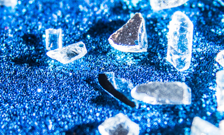
Imagine a 70-nanometer diamond particle—thousands of times smaller than a human hair—successfully entering a living cell and directly “sensing” the presence of free radicals inside it. This is the essence of the doctoral research conducted by drg. Aryan Morita, M.Sc., Ph.D., a faculty member in the Department of Dental Biomedicine at the Faculty of Dentistry, Universitas Gadjah Mada. He completed the research at the University of Groningen in the Netherlands in November 2020. The research addressed a fundamental question in cell biology: how can free radicals be measured directly inside a living cell without damaging it? The answer, remarkably, lies within the crystal structure of diamond.
Free Radicals and an Unresolved Problem
Free radicals are molecules with unpaired electrons that are naturally produced during cellular metabolism, particularly in the mitochondria. At balanced levels, they play important roles in normal cellular functions. However, when their levels become excessive or the systems responsible for eliminating them are impaired, a condition known as oxidative stress occurs. Oxidative stress has been associated with various degenerative diseases, cancer, cardiovascular disease, and the aging process itself.
The problem is that free radicals are extremely difficult to measure. They occur at low concentrations, have very short lifespans, and existing detection methods have significant limitations. For example, organic fluorescent dyes commonly used for detection are consumed when they react with free radicals, meaning they can only record the cell’s “past” rather than its current state. Conventional MRI techniques, meanwhile, require very large sample volumes and cannot image individual cells.
“With this technique, we can measure the concentration of free radicals in a living cell directly, in a single cell, and at that very moment, rather than obtaining a historical record as with other methods.” — drg. Aryan Morita, M.Sc., Ph.D.
Diamond as a Quantum Sensor Inside Cells
The solution developed by Aryan Morita is based on diamond magnetometry, a technique that exploits unique structural defects in diamond crystals known as nitrogen-vacancy (NV) centers. These defects form when a nitrogen atom replaces a carbon atom in the diamond crystal lattice, creating a “center” that is sensitive to magnetic signals in its surroundings. Because free radicals contain unpaired electrons that generate tiny magnetic fields, NV centers can “sense” them and convert these signals into measurable light.
The fluorescent nanodiamonds (FNDs) used in this research had a diameter of 70 nanometers and contained approximately 300–500 NV centers per particle. Their exceptional fluorescent stability, unlike that of organic dyes that fade over time, makes them ideal as long-term biological labels.
The first challenge was getting the particles into the cells. The yeast model organism, Saccharomyces cerevisiae, has a thick cell wall that naturally prevents particle entry. The research team developed a spheroplasting protocol, in which the cell wall was enzymatically removed and the FNDs were coated with cationic lipids to facilitate fusion with the cell membrane. The particles successfully entered the cytosol of living cells without causing significant toxicity.
From Young Cells to Old Cells: A Measurable Trail of Free Radicals
After successfully introducing the particles into the cells, Aryan Morita moved on to a larger question: could this technology distinguish between different cellular states?
In a series of experiments forming the core of his dissertation, he compared four yeast strains with different metabolic profiles, including a wild-type strain and three knockout mutants: sod1Δ, tor1Δ, and pex19Δ. The results were remarkable: diamond magnetometry was able to distinguish the four strains based on their intracellular free-radical burden, something conventional methods could not simultaneously achieve.
Furthermore, by following the same cells throughout 24 hours of chronological aging, the study confirmed that older cells had a higher free-radical burden than younger cells, consistent with the free radical theory of aging. Interestingly, the tor1Δ and pex19Δ strains, which are known to have longer lifespans, exhibited lower free-radical burdens. This finding provides a potential mechanistic explanation for their extended longevity.
The research also successfully detected the effects of antioxidants. Treatment with L-ascorbic acid (vitamin C) significantly increased T1 values, indicating a reduction in free-radical concentrations. This represented the first direct recording of antioxidant-induced free-radical reduction in a living cell at the single-cell level.
Relevance to Dental and Biomedical Science
This research is more than an achievement in quantum physics. Free radicals and oxidative stress play central roles in various pathological conditions relevant to dentistry, ranging from periodontal inflammation and dental pulp cell damage caused by irritation to aging processes in the oral mucosa. The ability to measure free-radical burden directly and in real time at the single-cell level opens opportunities for evaluating drugs, investigating antioxidant mechanisms, and gaining a deeper understanding of cellular responses to various agents.
In addition, this diamond nanosensor platform is modular. In another part of his dissertation, Aryan Morita successfully targeted diamond particles to the nucleus of yeast cells using antibodies specific to the nuclear pore complex, achieving a targeting success rate of 70%, compared with only 20% for non-targeted particles.
This approach could pave the way for measuring free radicals at specific locations within cells, including near DNA, which is important for understanding genetic damage caused by oxidative stress. Funded by an LPDP scholarship from the Ministry of Finance of the Republic of Indonesia, this research demonstrates that Indonesian scientists can contribute to the forefront of materials science and cell biology. Now that drg. Aryan Morita has returned to the Faculty of Dentistry at Universitas Gadjah Mada, expertise in quantum nanosensors has become part of the academic environment in Yogyakarta, bringing with it new and unanswered questions about life at the cellular level.
Authors: Achmad Zam Zam Aghasy, DDS, M.Kes.; Hazra Alifia Muharam
Photo: Freepik
Suber DOI: https://doi.org/10.33612/diss.136220127