
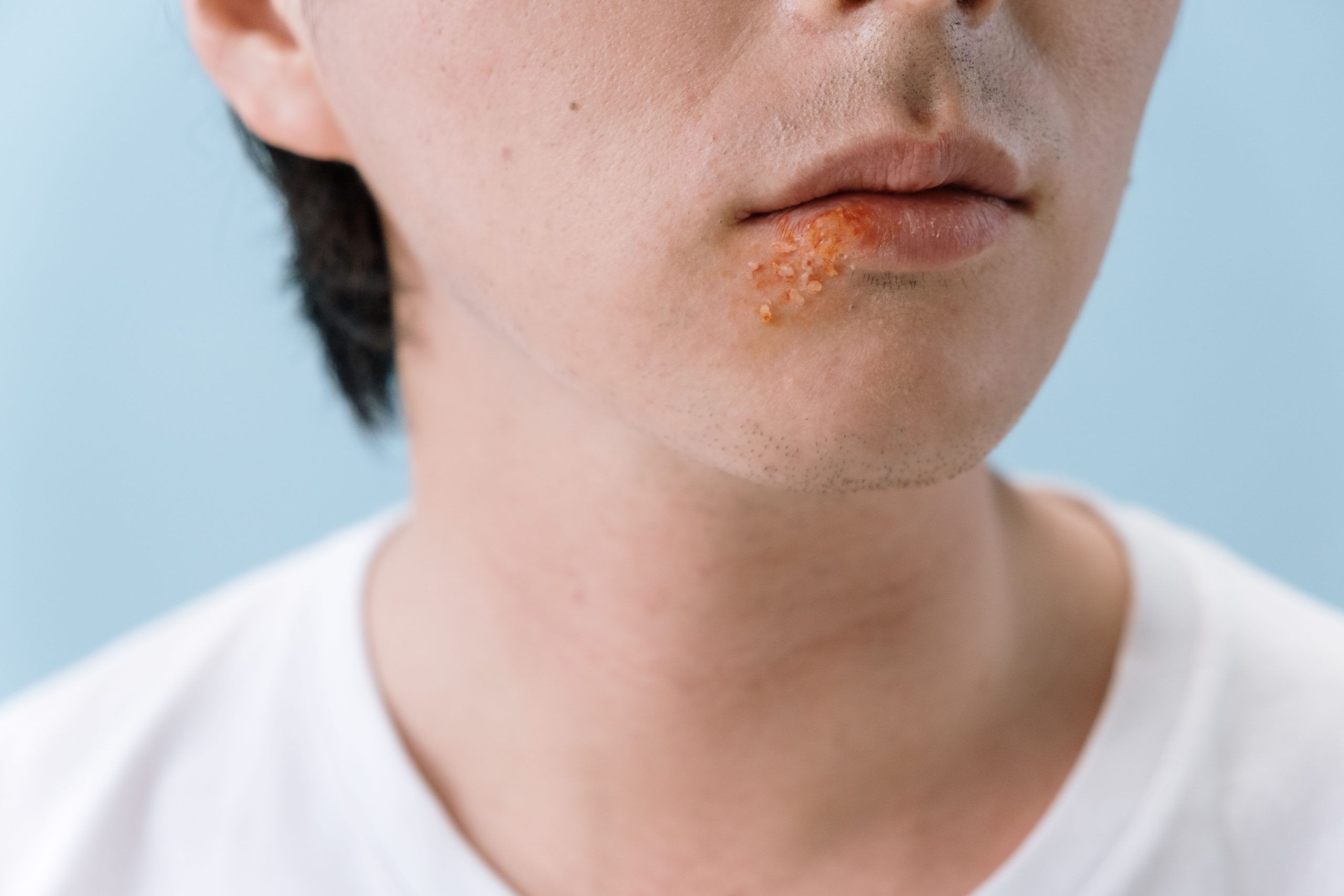
Imagine a child who underwent cleft lip and palate repair surgery as an infant, grew older, and later visited an orthodontic clinic with an upper jaw that was too narrow. The orthodontist decided to install an expansion device. However, something invisible was working against the treatment: scar tissue left behind from corrective surgery performed years earlier.
This was the starting point of research conducted by Rizqi Nurarifka Walupi under the supervision of Dr. drg. Cendrawasih Andusyana Farmasyanti, M.Kes., Sp.Ort(K), from the Department of Orthodontics, Faculty of Dentistry, Universitas Gadjah Mada. The study sought to answer a question that has rarely been quantitatively measured: to what extent does scar tissue actually inhibit maxillary movement during expansion procedures?
Narrow Jaw, Advanced Technology, and a Hidden Obstacle
Unilateral cleft lip and palate (UCLP) is one of the most common craniofacial malformations. It is characterized by a discontinuity of tissue that divides the maxilla into two segments: the major and minor segments. Patients with UCLP who have undergone corrective surgery often experience maxillary hypoplasia, a condition in which the upper jaw does not develop optimally.
To correct this condition, orthodontists use a device called a miniscrew-assisted rapid palatal expander (MARPE). Unlike conventional expanders that rely on teeth as anchorage points, MARPE is directly anchored into the palatal bone using mini-screws. This makes MARPE a suitable option for UCLP patients, whose periodontal condition is often insufficient to withstand the forces generated during expansion.
However, after corrective UCLP surgery, scar tissue forms in the palate and upper lip. This tissue is not passive. It creates tension, resists movement, and generates forces opposite to the expansion force produced by MARPE. Clinically, this phenomenon has long been recognized. However, the magnitude of its effect on jaw movement had never been measured using a controlled and quantitative approach.
Two Simulation Scenarios, One Important Finding
The research team developed a three-dimensional maxillary model complete with scar tissue, reconstructed from cone-beam computed tomography (CBCT) data of a UCLP patient. A digital MARPE model was also constructed. All components were integrated into a finite element model, a computational method capable of simulating force distribution and movement in complex structures such as the jawbone.
Two simulation scenarios were performed: First simulation: MARPE expansion forces were applied without considering scar tissue resistance. Second simulation: MARPE expansion forces were applied together with resistance forces generated by scar tissue in the palate and upper lip.
Movement was evaluated at sixteen reference points in the palatal and dental regions across three axes: x-axis: lateral movement, y-axis: vertical movement, and z-axis: anteroposterior movement.
The results were consistent across both simulations. The maxilla moved laterally along the x-axis, downward along the y-axis, and forward along the z-axis. The movement pattern showed that the minor segment consistently moved farther than the major segment along the x and z axes, with movement decreasing from the anterior to posterior regions. Conversely, the major segment showed greater movement along the y-axis.
“The average movement values in the second simulation were lower than those in the first simulation across all three axes,” according to the main finding of the study. The conclusion was clear: scar tissue does not change the pattern of maxillary movement, but it significantly reduces the magnitude of movement achieved.
The Numbers Behind the Scar Tissue
This finding has important clinical implications. Orthodontists treating UCLP patients with MARPE may already suspect that scar tissue creates resistance during expansion. However, without quantitative data, it has been difficult to predict how much correction is required or whether the targeted expansion can be achieved according to the treatment plan.
This study provides simulation-based evidence that scar tissue resistance is a real variable affecting expansion outcomes. The fact that movement patterns remained the same in both simulations provides encouraging information: the direction of correction is not distorted by scar tissue. However, the reduction in movement magnitude reminds clinicians that treatment planning for UCLP patients must account for this factor.
The finite element analysis simulation method used in this research also opens opportunities for future studies. Computational models allow researchers to test scenarios that cannot easily be performed directly on patients, including variations in scar tissue thickness, miniscrew placement, and different expansion forces.
For UCLP patients who have carried the physical consequences of surgery since childhood, research like this represents more than numbers and simulations. It is a scientific effort to ensure that their treatment is carefully planned by considering every factor within the oral cavity—including scars that may not be visible but have a real impact on treatment outcomes.
Authors: Nanda Ayu, drg. Achmad Zam Zam Aghasy, M.Kes.
Photo: Pexels