
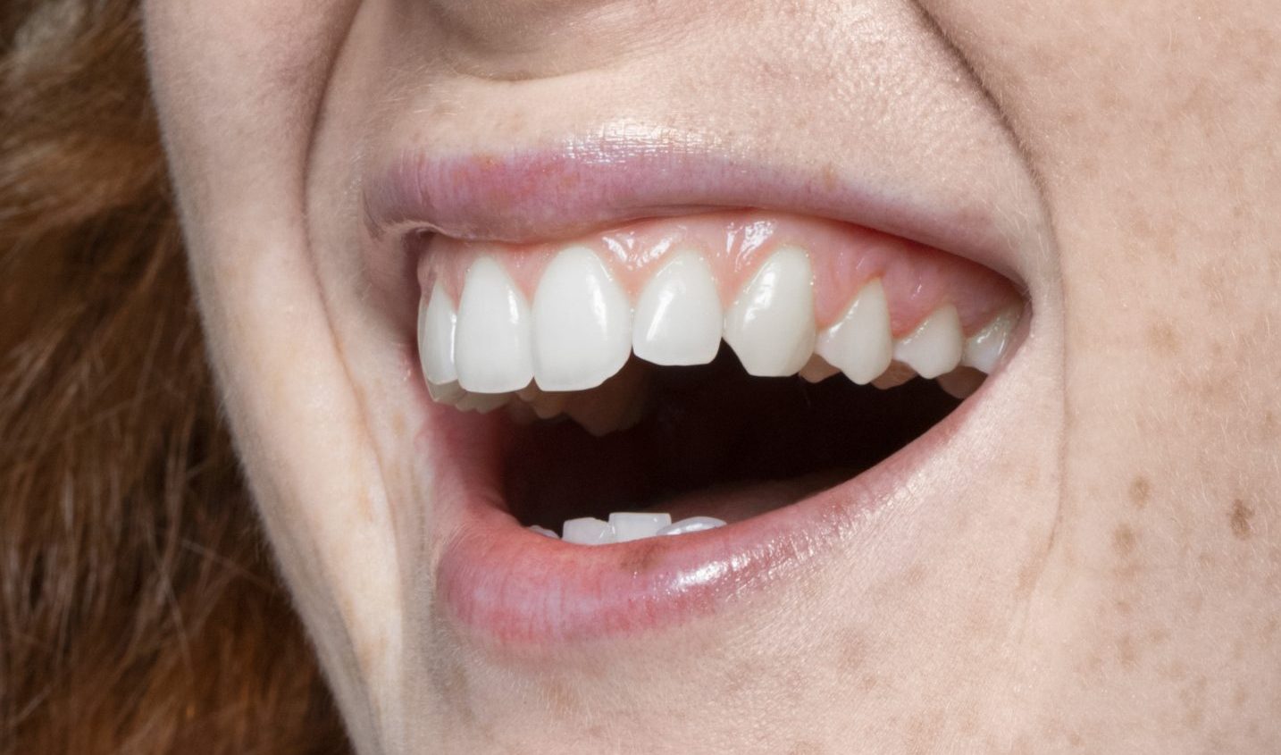
Imagine gum tissue that should firmly support the teeth slowly breaking down from within. Not because of an accident or a tumor, but because of a chain reaction triggered by a single type of bacterium. This is what drg. Dyah Listyarifah, M.Sc., D.Med.Sci., a researcher from the Department of Biomedical Dentistry, Faculty of Dentistry, Universitas Gadjah Mada, and her international research team discovered. Their immunohistological study, published in an international scientific journal, revealed how the bacterium Treponema denticola triggers cell death, intensifies inflammation, and paves the way for more extensive periodontal tissue destruction—a process that has not yet been fully understood.
Treponema denticola: An Invader That Penetrates Deep into Tissue
Periodontitis is an inflammatory disease affecting the tissues that support the teeth and, if left untreated, can eventually lead to tooth loss. It is not merely a matter of poor oral hygiene. Beneath the inflammation visible on the surface, a biological battle is taking place at the cellular level.
In this study, the team, which included experts from the University of Helsinki, Karolinska Institutet in Sweden, and Université Laval in Canada, analyzed gum tissue samples from two groups of patients: 10 patients with periodontitis and five patients with gingivitis, a milder form of gum inflammation. Each sample was examined using immunohistochemical and immunofluorescence staining techniques, methods that allow researchers to directly trace specific proteins and cells within tissue.
The results were striking. Treponema denticola was found in every periodontitis tissue sample without exception. The bacterium penetrated the sulcular epithelium, the layer of cells lining the space between the gums and teeth, and extended deeper into the underlying connective tissue known as the lamina propria. The bacterium's proteolytic enzyme, known as chymotrypsin-like proteinase (CTLP), was detected not only inside cells but also in the extracellular space—a sign that the bacterium had truly invaded the tissue.
Dead Cells Can Actually Worsen Inflammation
One of the most intriguing findings of the study concerned apoptosis, or programmed cell death. Under normal conditions, apoptosis is a mechanism the body uses to remove damaged cells in an orderly and controlled manner. In periodontitis, however, this process appears to become fuel for inflammation.
The researchers measured levels of caspase-3, an active marker of apoptosis, and found significantly higher levels in periodontitis tissue than in gingivitis tissue. Additional confirmation came from TUNEL staining, a technique used to detect DNA fragmentation, a hallmark of dying cells. These apoptotic cells accumulated particularly in the junctional epithelium, the area where the gums attach to the teeth and which serves as a major entry point for bacteria.
More concerningly, macrophages—the immune cells responsible for engulfing dead cells to prevent them from triggering inflammation—also underwent apoptosis in large numbers. As a result, dead cells that should have been cleared accumulated, lost their membrane integrity, and eventually ruptured, releasing their cellular contents into surrounding tissue, including harmful molecules known as damage-associated molecular patterns (DAMPs).
Danger Signals That Intensify the Inflammatory Cycle
This is where HMGB1 (High Mobility Group Box 1) becomes crucial. Under normal conditions, HMGB1 is a protein that functions inside the cell nucleus and contributes to genetic transcription. However, when cells experience severe stress or die, HMGB1 moves from the nucleus into the cytoplasm and is eventually released into the extracellular space, where it takes on a new role as a danger signal that triggers an immune response.
In periodontitis tissue, the researchers observed a clear shift in HMGB1 expression. In gingivitis, the protein remained predominantly localized in the cell nucleus. In periodontitis, however, it was found extensively in the cytoplasm and even outside the cells. Cytoplasmic HMGB1 expression was statistically higher in the periodontitis group than in the gingivitis group.
“The presence of Treponema denticola (particularly CTLP), apoptosis, HMGB1, and inflammatory markers suggests their potential involvement in the pathogenesis of periodontitis.”
The released HMGB1 then binds to its receptors, particularly TLR4 (Toll-like receptor 4), whose expression was also significantly increased in periodontitis tissue. The binding of HMGB1 to TLR4 stimulates immune cells to produce pro-inflammatory cytokines, namely IL-1β and IL-8—two molecules known to contribute to soft tissue and bone destruction in periodontitis. This creates a self-perpetuating cycle: the more cells die, the more HMGB1 is released; the more HMGB1 is released, the stronger the inflammation becomes; and the stronger the inflammation, the more cells ultimately die.
Implications for the Diagnosis and Treatment of Periodontitis
The study has several limitations, including its relatively small sample size and considerable age differences between the periodontitis and gingivitis groups. The researchers themselves acknowledged that further studies involving larger samples, including in vitro studies and animal experiments, are needed to establish this mechanism in greater detail.
Nevertheless, the findings open up an important new perspective. Treatment of periodontitis has traditionally focused on eliminating bacteria through procedures such as scaling and root planing, antibiotics, or periodontal surgery. This study suggests that disrupting HMGB1 signaling or preventing excessive accumulation of apoptotic cells could become a promising additional therapeutic strategy.
Periodontitis is not merely a gum disease. It is a complex inflammatory ecosystem in which bacteria, dying cells, and signaling molecules work together to worsen tissue destruction. Understanding how this ecosystem operates is the first step toward truly stopping it.
Authors: Achmad Zam Zam Aghasy, DDS, M.Kes.; Hazra Alifia Muharam
Photo: Freepik