
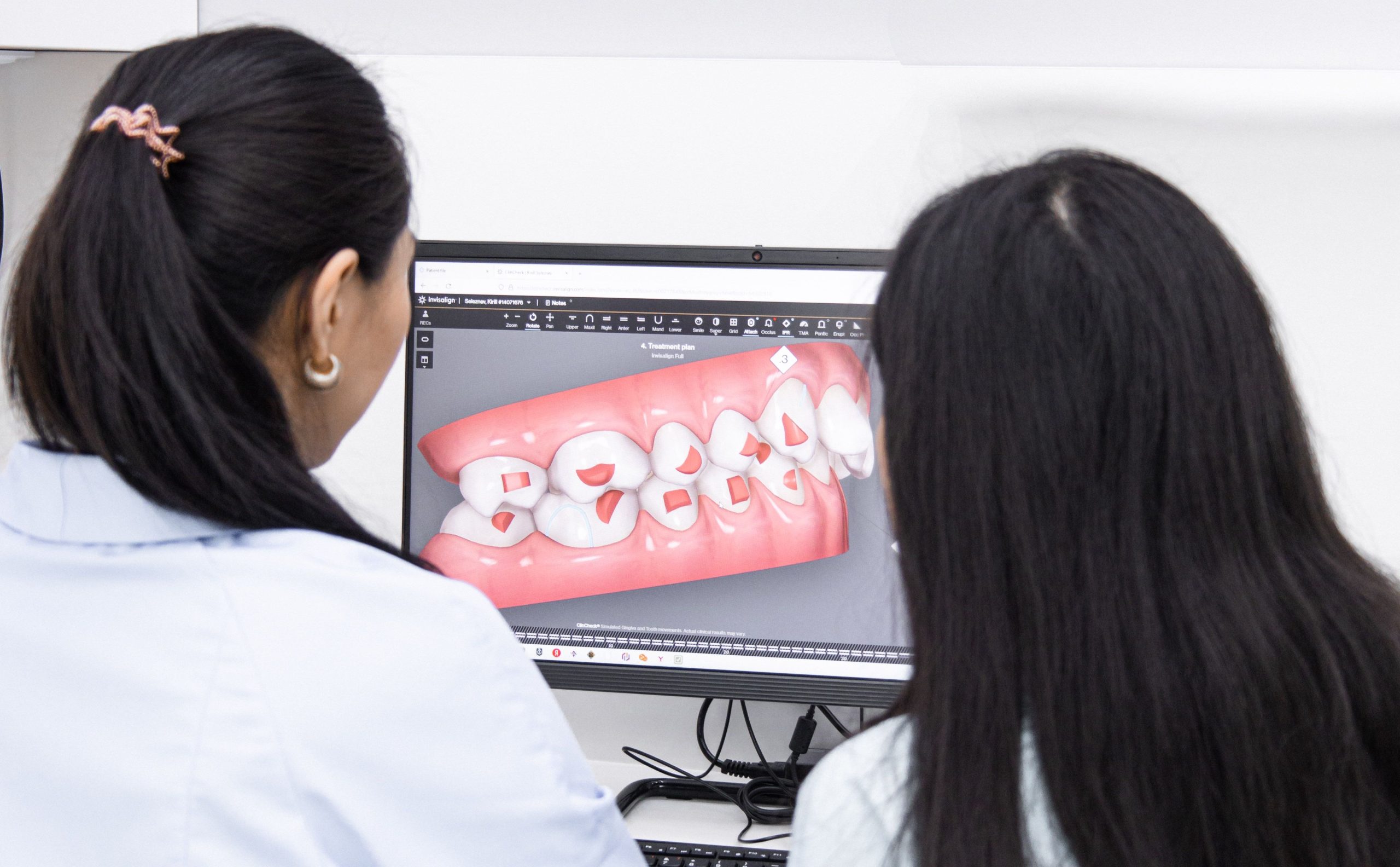
A fundamental question has emerged among orthodontic clinics transitioning toward digital technology: can three-dimensional dental models displayed on a computer screen be trusted to be as accurate as conventional plaster models that have served as the gold standard for decades? A study involving researchers from the Department of Orthodontics, Faculty of Dentistry, Universitas Gadjah Mada (UGM), and Universitas Indonesia attempted to answer this question systematically.
The results showed that all four tested measurement methods, including conventional plaster models, 2D digital scanning, 3D intraoral scanning, and Cone Beam Computed Tomography (CBCT), were proven to be accurate. However, their levels of accuracy were not entirely equivalent.
When Plaster Models Remain the Gold Standard
Study models serve as the foundation of orthodontic diagnosis. Through these models, orthodontists measure the mesiodistal width of teeth, calculate space discrepancies, and plan tooth movements that may take years to complete. Even a one-millimeter measurement error can determine the difference between a successful treatment plan and an inaccurate one.
The study, published in the April 2023 edition of the Journal of Dentomaxillofacial Science, was designed as an experimental comparative study. The research team, including Dr. drg. Dyah Kurnia, Sp.Ort. from the Faculty of Dentistry UGM, used a typodont as the control group or “gold standard”—a dental model with complete and standardized morphology. Using the same typodont, five groups of models were created using different methods: conventional plaster models, 2D scanning using a flatbed scanner, 3D intraoral scanning using the Medit i-500 device, and CBCT imaging using the Orthopantomograph OP300.
Each measurement was repeated three times by the same observer and then averaged. The measured parameters included the total mesiodistal tooth width and the interpremolar distance.
The results showed that conventional plaster models achieved the highest correlation values compared with other methods. For measuring total mesiodistal tooth width, plaster models achieved an R value of 0.982, while 3D intraoral scanning produced R = 0.961, CBCT produced R = 0.941, and 2D scanning produced R = 0.913. All values exceeded the statistical significance threshold (p < 0.05).
Why Plaster Models Perform Better, and the Limitations of Digital Methods
The superiority of plaster models can be explained logically. Their measurement process uses the same digital calipers applied to measure the typodont control model, resulting in highly similar measurement conditions. Although shrinkage of alginate impression materials and gypsum degradation may cause minor discrepancies, these differences are not significant enough to substantially reduce accuracy.
Nevertheless, plaster models have unavoidable disadvantages: they require physical storage space, are vulnerable to damage, and cannot be instantly shared with colleagues or other specialists.
This is where digital technology provides clear advantages. Three-dimensional intraoral scanning, for example, uses confocal imaging technology, which can detect object surface details and calculate distances based on lens focal length. The results are stored in an .stl file format that can be accessed from anywhere, eliminating the need for physical storage and reducing the risk of model damage. For patients with strong gag reflexes or limited mouth opening ability, this method is also considerably more comfortable than conventional alginate impression procedures.
“The three-dimensional model has many advantages over conventional plaster models in orthodontic aspects, including it is easier to share with doctors or other professionals from anywhere for advice on treatment plans or diagnostics.”
In contrast, 2D scanning was the least accurate method, particularly for measuring interpremolar distance (R = 0.881). The convex structure of posterior teeth and inconsistent scanning angles make anatomical reference points—especially the mesial grooves on the occlusal surfaces of premolars—difficult to identify precisely.
CBCT: Advanced Technology but Not Suitable for Every Case
CBCT measurements demonstrated very strong accuracy, with R values above 0.90 for both parameters. This method uses a segmentation system that allows clear visualization of individual tooth contours, enabling highly accurate mesiodistal width measurements from an occlusal view.
However, CBCT also has limitations. It is expensive, requires additional operator training, and exposes patients to higher radiation doses compared with conventional radiography. In clinical practice, CBCT is primarily indicated for specific cases such as impacted tooth evaluation, pharyngeal airway assessment, mini-implant planning, or preparation for orthognathic surgery. Using CBCT solely for routine model analysis would be excessive, both in terms of cost and radiation exposure risks.
Choosing a Method Depends on Clinical Considerations
The findings of this study do not indicate that one method is absolutely superior to another. All methods are accurate. The difference lies in their clinical context and application.
Clinics with limited access to digital technology can continue relying on conventional plaster models while maintaining scientifically acceptable diagnostic accuracy. Clinics seeking improved storage efficiency and easier data sharing between clinicians can adopt 3D intraoral scanning without significantly compromising accuracy. Meanwhile, CBCT remains relevant for specific clinical indications requiring comprehensive three-dimensional visualization.
This study received ethical approval from the Health Research Ethics Committee of Universitas Gadjah Mada. The research was conducted using facilities from the Department of Orthodontics, Faculty of Dentistry UGM, and the Radiology Department of Prof. Soedomo Dental Hospital, Yogyakarta.
On one hand, these findings provide reassurance: transitioning into the digital era does not necessarily mean sacrificing diagnostic precision. On the other hand, they serve as a reminder that advanced technology does not automatically replace deep understanding of dental anatomy and the precision of clinical measurement.
Authors: Nanda Ayu, drg. Achmad Zam Zam Aghasy, M.Kes.
Photo: Pexels