
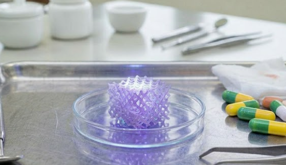
Imagine a thin sheet, only about one hundred micrometers thick, that could be implanted in an area of jawbone damaged by periodontitis. This sheet is not merely a passive support. It releases calcium minerals in a controlled manner, allows bone cells to attach, and remains stable long enough for new tissue to form—then gradually degrades on its own. This is what Dr. drg. Retno Ardhani, M.Sc., from the Department of Dental Biomedical Sciences, Faculty of Dentistry, Universitas Gadjah Mada, together with a multidisciplinary research team from Indonesia and Germany, sought to develop in a study published in the International Journal of Polymer Analysis and Characterization in 2022. The research addressed a fundamental question: how can the properties of a biological scaffold be precisely controlled using ultraviolet light as the trigger?
Why Gelatin Scaffolds Alone Are Not Enough
In guided bone regeneration (GBR) procedures, a technique used to regenerate bone in severe cases of periodontitis, dentists need a barrier membrane that can direct the growth of new bone while preventing soft tissue from filling the defect prematurely. Gelatin, a natural protein derived from collagen, has long been used as a scaffold material because it is readily biodegradable and supports cell attachment. However, gelatin has a significant drawback: it degrades too quickly and is mechanically too weak to withstand the dynamic environment of the oral cavity.
Various modification strategies have previously been explored, including blending gelatin with other polymers, using carbodiimide-based chemical reactions, and applying light irradiation. However, many conventional chemical methods require two reactive components to approach each other and react, a process that becomes considerably slower when the material is in a dry and solid state. This is where the research team identified an opportunity that had not been fully explored.
C,H-Insertion Reaction: UV Light as a Molecular Adhesive
The strategy developed by Dr. drg. Retno Ardhani, M.Sc., and her team was based on the use of a C,H-insertion reaction, a chemical mechanism in which benzophenone groups exposed to UV light “insert” themselves into nearby C–H bonds, forming new covalent bonds without requiring a specific reactive partner. Because gelatin, as a polypeptide, contains abundant C–H bonds, it serves as an ideal substrate for this reaction.
The team synthesized a series of photoreactive polymers known as P(DMAA-n%MABP-OH), copolymers of N,N-dimethylacrylamide (DMAA) and 2-hydroxy-(4-methacryloyloxybenzophenone), with benzophenone contents of 1%, 5%, and 10%. These polymers were then applied to a CHA/gelatin scaffold—a composite consisting of carbonated hydroxyapatite (CHA), a mineral resembling the inorganic component of natural bone, and gelatin—through a brief immersion process followed by UV irradiation. The process produced a PDMAA-based hydrogel that became covalently attached to the scaffold rather than merely adhering to its surface.
The chemical structures of the polymers were confirmed using nuclear magnetic resonance (NMR) spectroscopy, while the reactivity of benzophenone under UV exposure was monitored using UV-Vis spectroscopy. The results showed a gradual decrease in benzophenone absorbance with increasing irradiation time, confirming that the photochemical reaction occurred and that new C–C bonds were formed.
Properties That Can Be Tuned, Not Merely Improved
One of the most interesting findings of the study was the ability to tune the properties of the scaffold simply by altering the benzophenone content of the photoreactive polymer. As the n value, representing the MABP-OH content, increased, the density of crosslinking also increased, resulting in greater changes in the scaffold's properties.
From a mechanical perspective, the scaffold coated with P(DMAA-10%MABP-OH) demonstrated the highest wet tensile strength and elastic modulus among the groups, highlighting the role of crosslink density in improving mechanical durability. In terms of degradation, the uncoated scaffold completely degraded after 72 hours in PBS solution and 24 hours in 0.1 N HCl. In contrast, the scaffold coated with P(DMAA-10%MABP-OH) remained stable for approximately 1,536 hours, or around 64 days, under acidic conditions designed to simulate an inflammatory environment.
The release of calcium ions (Ca²⁺) from CHA, which are important for stimulating osteoblast proliferation and bone tissue formation, could also be controlled. Polymer-coated scaffolds released Ca²⁺ more slowly and consistently than uncoated scaffolds, suggesting the potential to adjust the rate of mineral release according to clinical requirements.
“This dual strategy, in which the material’s interior is modified while its surface is simultaneously coated in a single process, enables the tuning of scaffold properties as well as the control of active compound release.” — Retno Ardhani et al., International Journal of Polymer Analysis and Characterization, 2022
The swelling index, which reflects the scaffold’s ability to absorb fluid and expand in a manner resembling the extracellular matrix environment, also demonstrated a controllable pattern. The scaffold containing P(DMAA-5%MABP-OH) achieved the highest swelling index, reaching nearly seven times its initial mass, and surprisingly demonstrated the best cell adhesion, almost twice that of the uncoated scaffold.
Living Cells, Attached Cells: Biocompatibility Testing
A critical question in the development of implantable materials is whether the material is safe for living cells. The research team evaluated cytotoxicity using primary pericoronal cells, which are cells associated with the crowns of unerupted teeth and were obtained from the Department of Oral Medicine, Faculty of Dentistry, Universitas Gadjah Mada. All scaffold variants, both coated and uncoated, demonstrated cell viability above 80%. This exceeded the safety threshold specified by ISO 10993-5, indicating that the materials were not cytotoxic.
Cell attachment was evaluated using MC3T3E1 preosteoblast cells. The addition of the photoreactive polymer coating increased cell adhesion to the scaffold, with the P(DMAA-5%MABP-OH) scaffold producing the highest cell-adhesion value. This phenomenon was associated with the ability of the swelling hydrogel to prevent nonspecific protein adsorption while facilitating specific interactions between RGD peptide sequences in gelatin and cell receptors. The CHA mineral also contributed by providing calcium and phosphorus required for bone cell growth.
This research demonstrates that combining CHA as a bioactive inorganic component, gelatin as a natural polymer that supports cell attachment, and PDMAA as a biopassive synthetic polymer can create a multifunctional scaffold with promising potential for GBR applications. The result is not simply a scaffold that is “better,” but one whose properties can be designed from the outset according to specific requirements—with light serving as the tool for controlling those properties.
Authors: Achmad Zam Zam Aghasy, DDS, M.Kes.; Hazra Alifia Muharam
Photo: Freepik
DOI: https://doi.org/10.1080/1023666X.2022.2076012