
Millions of people worldwide suffer from bone damage every year due to accidents, tumors, or bone diseases. The problem is not merely the pain, but also the limited availability of bone replacement materials that are truly compatible with the human body. Dr. drg. Anne Handrini Dewi, M.Kes., a researcher from the Department of Biomedical Dentistry, Faculty of Dentistry, Universitas Gadjah Mada, is attempting to address this challenge through research published in The Indonesian Journal of Dental Research. The study tested a combination of two natural materials, carbonate apatite and chitosan, as a bone substitute in an animal model. The results were quite remarkable: the combination of the two materials was shown to significantly promote new bone formation.
Artificial Bone from Materials Found in Nature
Carbonate apatite is not foreign to the human body. In fact, this mineral is a major component of our bones and teeth. Unlike hydroxyapatite, which has traditionally been widely used as a bone substitute, carbonate apatite more closely resembles the composition of natural human bone, making it easier for the body's cells to absorb and break down.
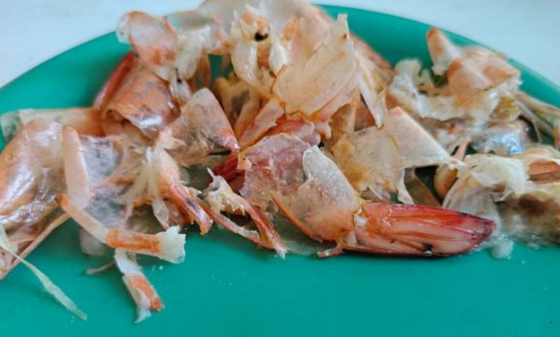
Chitosan, on the other hand, is a natural polymer derived from chitin, a substance that forms the shells of shrimp and crabs as well as the exoskeletons of insects. In the medical field, chitosan has long been recognized for its tissue-friendly properties: it is non-toxic, biodegradable, has antibacterial effects, and can support wound healing.
Dr. Anne combined the two materials in a 1:1 ratio to form a paste that could be directly applied to bone defects. The study was conducted at the Integrated Research and Testing Laboratory (LPPT) of UGM using 18 three-month-old male Sprague Dawley rats weighing 250–300 grams. A 3 × 1.25 × 1 mm³ defect was created in the tibia of each rat using a low-speed dental bur and then filled with the test material. Observations were made during the first, second, and third weeks after implantation.
Bone-Forming Cells Work Faster
The results provided an interesting picture. During the first week, the groups treated with carbonate apatite and the carbonate apatite–chitosan combination showed higher numbers of osteoblasts (bone-forming cells) and osteoclasts (cells responsible for remodeling old bone) than the control group, which received no material. This indicated that the materials immediately stimulated bone regeneration activity from the early stages.
However, during the second and third weeks, the pattern was reversed. The number of osteoblasts in the treatment groups decreased, while remaining high in the control group. This was not a negative finding; in fact, the opposite was true.
"The decrease in the number of osteoblasts during the third week indicates that osteoblast calcification into osteocytes occurred more rapidly in the treatment groups, meaning that the bone had entered the maturation phase earlier," wrote Dr. Anne Handrini Dewi, M.Kes., and Dr. drg. Andi Triawan, Sp.Ort(K), in the paper.
In other words, the bone in the groups receiving the test materials had already "passed" the formation phase and was progressing toward maturation, while the control group was still working to fill the defect. Light microscopy observations during the third week showed that the carbonate apatite and carbonate apatite–chitosan groups exhibited more mature bone than the control group.
Chitosan Dissolves More Slowly but Remains Promising
Another interesting finding concerned the rate at which the materials were absorbed. Under microscopic examination, residual chitosan was still visible during the first and second weeks, indicating that chitosan was absorbed more slowly than pure carbonate apatite. As a result, carbonate apatite without chitosan was shown to be more effective in increasing the number of osteoblasts and osteoclasts, particularly during the first week.
Nevertheless, the combination of the two materials remains promising because the versatility of chitosan allows it to be formulated as powder, paste, film, or fiber, depending on clinical requirements. Chitosan can also be combined with various other biomaterials, such as hydroxyapatite, hyaluronic acid, and growth factors.
The study concluded that the combination of carbonate apatite and chitosan has the potential to serve as a biocompatible bone substitute candidate. Going forward, Dr. Anne suggested modifying chitosan powder into nanoparticles to optimize its absorption in bodily fluids.
The remaining question is: to what extent can findings from this animal model be translated into clinical applications in humans? The road ahead is still long, but the scientific foundation is now stronger.
Authors: Achmad Zam Zam Aghasy, DDS, M.Kes.; Hazra Alifia Muharam
Photo: Pixabay