
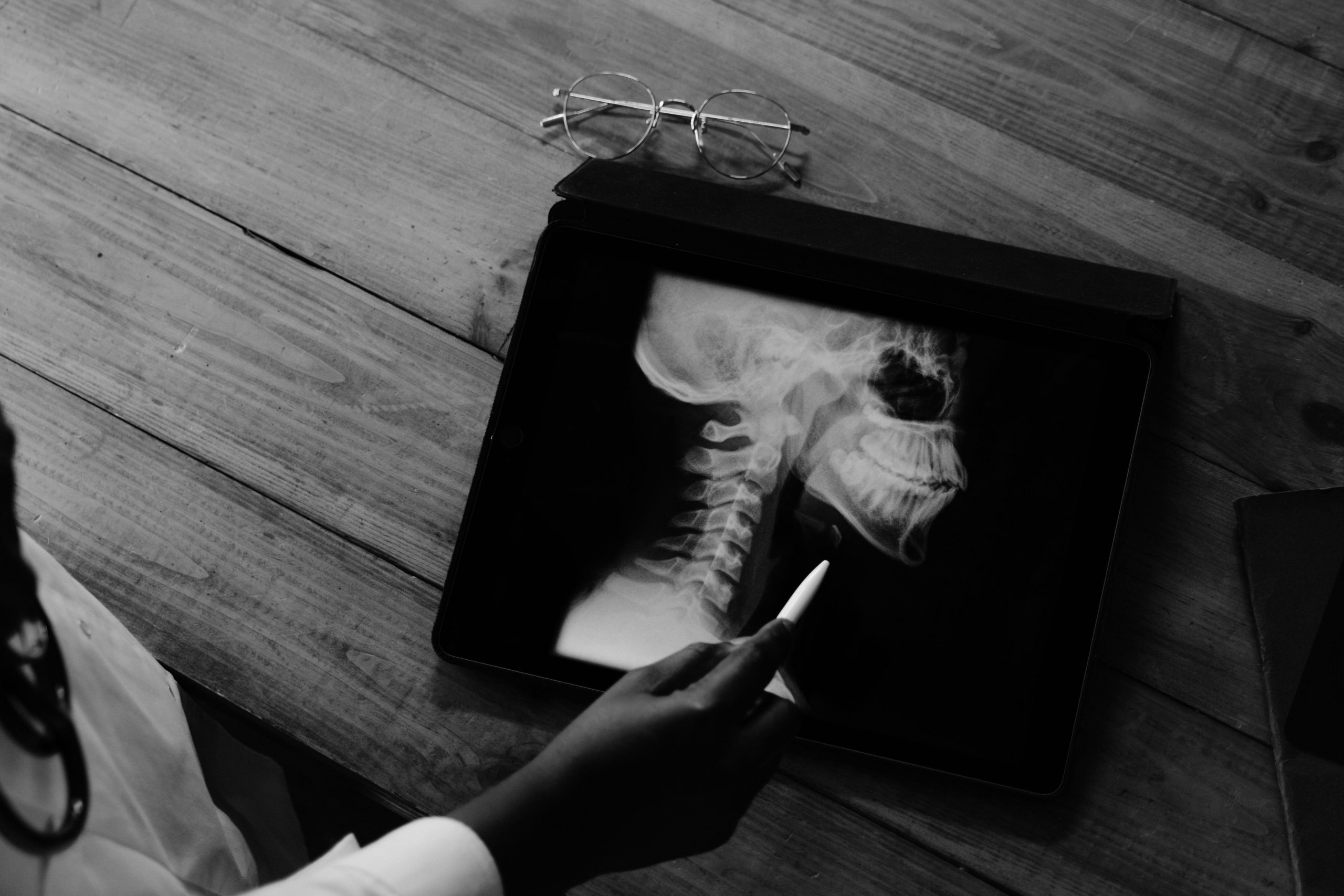
Osteoporosis is often referred to as a “silent disease.” It gradually weakens the bones without symptoms, pain, or noticeable warning signs—until one day a fracture occurs. In postmenopausal women, the risk is even greater: declining estrogen levels accelerate bone resorption, and damage can occur long before a person realizes that she is at risk. The question is: is there an earlier, simpler, and more affordable way to detect it?
A study involving drg. Silviana Farrah Diba, Sp. RKG. Subsp. RP (K) from the Department of Oral and Maxillofacial Radiology, Faculty of Dentistry, Universitas Gadjah Mada, sought to answer that question—not with a conventional bone scanner, but with a device already available in dental clinics: cone-beam computed tomography (CBCT).
The Jaw as a Mirror of Overall Bone Health
The study was published in the World Journal of Dentistry in 2020 and was led by Prof. Azhari from Universitas Padjadjaran, with drg. Silviana Farrah Diba serving as one of the principal researchers responsible for data acquisition, analysis, and interpretation.
A total of 17 postmenopausal women aged 50–84 years who visited the Universitas Padjadjaran Dental Hospital participated in the study. From these 17 subjects, the research team obtained 34 observation sites—two sites on the mandible from each subject—and analyzed them using high-resolution CBCT images.
The researchers measured not just one parameter, but six. For cortical bone, they used three indices based on the ratio of cortical thickness to specific distances from the mental foramen, an anatomical opening in the mandible through which a nerve emerges. For trabecular bone, they measured bone volume fraction (BV/TV), trabecular thickness (Tb.Th), and trabecular separation (Tb.Sp) using BoneJ software.
One day after the CBCT scan, the subjects underwent blood sampling following a 12-hour fast. From the blood samples, the researchers measured beta-crosslaps (β-CTx), a biochemical marker reflecting the rate at which the body breaks down type I collagen, the main component of the bone matrix. The higher the β-CTx level, the greater the rate of bone resorption.
Signals That Can Be Read from Three-Dimensional Images
The results were fairly convincing. Both trabecular and cortical parameters showed significant positive correlations with β-CTx levels. Trabecular parameters showed a correlation of r = 0.477 (p = 0.049), while cortical parameters showed a correlation of r = 0.411 (p = 0.038).
These figures may sound technical, but their meaning is straightforward: women with higher β-CTx levels—which indicate more active bone resorption—tended to have poorer trabecular and cortical conditions in the jaw, and these changes could be detected on CBCT images.
“Cone-beam computed tomography can be used to assess the quality of mandibular cortical and trabecular bone, which correlates with β-CTx levels in postmenopausal women, for the early detection of declining bone quality.”
This finding is important because CBCT is already commonly used in dental clinics for various purposes, ranging from implant planning to diagnosing jaw abnormalities. This means that an existing device could potentially provide additional information about osteoporosis risk without requiring a separate and more expensive examination.
Earlier Detection, Before the Bones Become Truly Fragile
The clinical implications of this study extend beyond academia. Dentists occupy a unique position: patients often visit them regularly, sometimes more frequently than they visit general practitioners, and radiographic imaging is already part of routine dental examinations. If changes in jawbone structure can serve as an early signal of osteoporosis, dental visits could become an opportunity for initial detection—before a fracture alerts patients to the presence of a serious problem.
The study also highlights the practical value of β-CTx as a biomarker. Unlike some other biochemical markers, β-CTx does not require creatinine correction and is not excreted by the kidneys, allowing it to more directly reflect bone matrix degradation without interference from certain external factors.
Of course, the study has limitations. The sample size was relatively small, consisting of only 17 participants, and the analytical software used could display image slices but could not fully reconstruct the three-dimensional trabecular structure. The researchers themselves emphasized the need for further studies involving larger samples and more advanced equipment.
Nevertheless, that is precisely where the relevance of this study lies: it is not the final word, but rather an invitation to take a more serious look at the role of dental radiology in the early detection of systemic diseases. The jaw, it turns out, is not just about teeth. It contains a story about the entire body—and CBCT may help us read it.
Source DOI: http://10.5005/jp-journals-10015-1704
Authors: Anny Anggraini; drg. Achmad Zam Zam Aghasy, M.Kes.
Photo: Pexels