
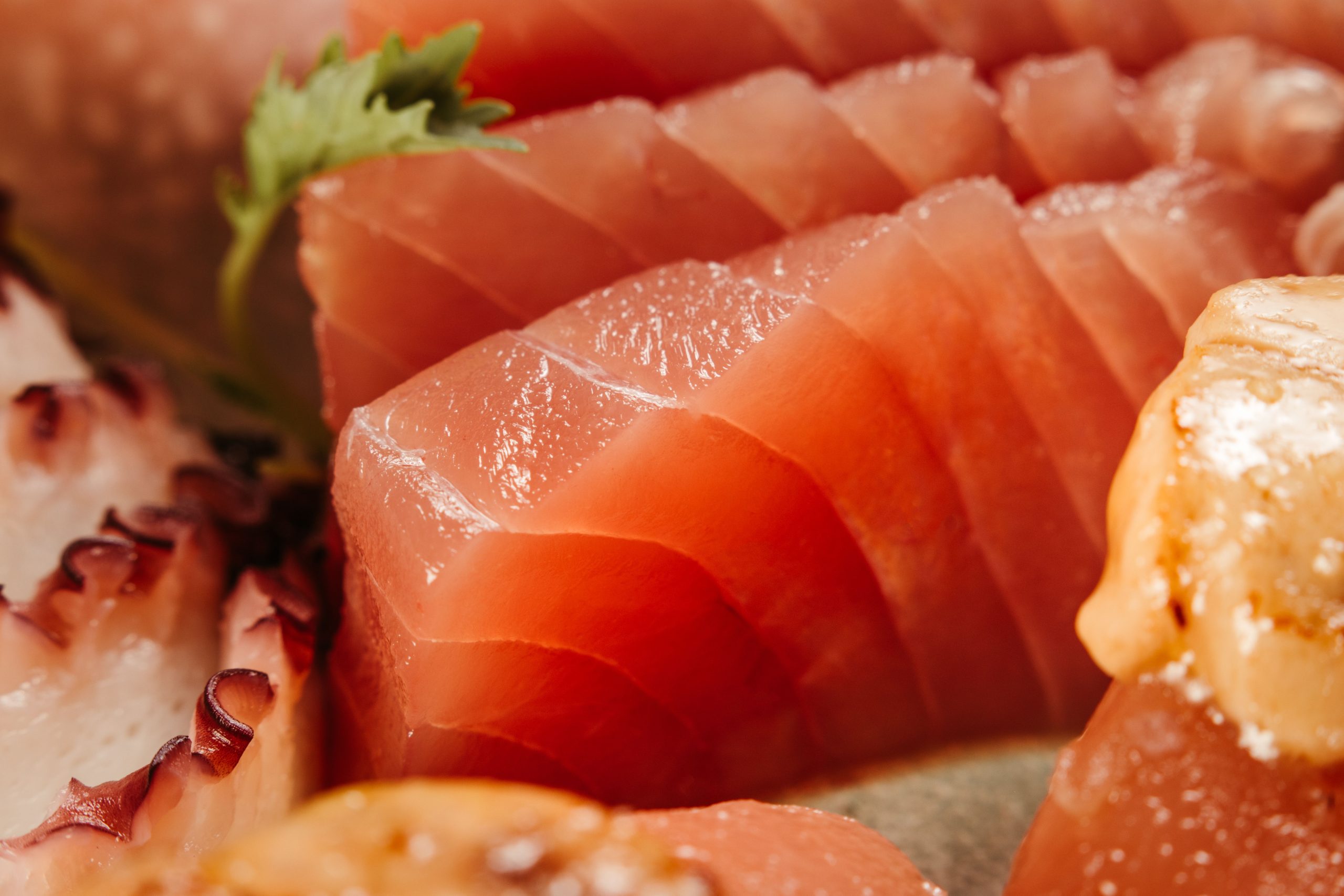
Waste that has traditionally ended up merely as an ingredient in fish feed may actually hold much greater potential. Yellowfin tuna bones (Thunnus albacares), which are usually discarded after the meat is removed, have now become a promising material for testing in the field of dental biomaterials. Researchers from the Department of Oral Biology, Faculty of Dentistry, Universitas Gadjah Mada, demonstrated that hydroxyapatite extracted from these fish bones is not only safe for cells but also promotes cell proliferation.
From Fish Processing to the Research Laboratory
Prof. drg. Tetiana Haniastuti, M.Kes., Ph.D., together with her research team, Ananda Mutiara Wening, Firda Arifatul Faqiha, and Dian Az Zahra, conducted a series of laboratory tests to evaluate the effect of tuna bone-derived hydroxyapatite (THA) on cell proliferation. The study was published in the Journal of International Dental and Medical Research in 2020.
The choice of yellowfin tuna was not without reason. This fish is among the most widely consumed species worldwide, yet its use has traditionally focused only on its meat. Its bones, which are rich in calcium and phosphorus, are instead discarded. This is where the research idea originated: what if this waste could be processed into a material capable of supporting human bone regeneration?
The production of THA began by heating the bones under a pressure of 80 kPa for 1.5 hours, followed by oven drying for eight hours per day over four consecutive days. The bones were then ground into a fine powder, sintered at 700°C for five hours, and sieved to obtain particles measuring 74 micrometers. The resulting THA powder was then ready to be tested on living cells.
Living Cells Respond Positively, Rather Than Showing Toxicity
The researchers used two cell lines commonly employed in biological research: Vero cells, derived from the kidneys of African green monkeys, and HeLa cells, derived from human cervical tumors. Both were selected because they have been established as reliable models for in vitro cytotoxicity testing. in vitro.
THA was administered at various concentrations, ranging from 12.5 to 100 μg/ml, and observed for 24 and 48 hours using the MTT assay. This method measures mitochondrial enzyme activity in living cells: the greater the number of active and healthy cells, the higher the measured absorbance value.
The results were striking. Rather than showing signs of toxicity, both cell types actually proliferated more than the control group. Vero cell proliferation reached 105.26–109.12% after 48 hours of exposure. Meanwhile, HeLa cells showed proliferation rates of 104.35–109.8%, with significant effects already observed after the first 24 hours.
“Vero and Hela cells enhance their proliferation after exposed to THA, suggesting the potency of THA to be used as a biomaterial.” — Prof. drg. Tetiana Haniastuti, M.Kes., Ph.D., et al., Journal of International Dental and Medical Research, 2020
According to ISO 10993-5 standards for the biological evaluation of medical devices, cell viability above 80% is categorized as non-cytotoxic. With proliferation values consistently exceeding 100%, THA passed this test by a considerable margin.
Why Is Natural Hydroxyapatite More Appealing?
Hydroxyapatite (HA), with the chemical formula Ca₁₀(PO₄)₆(OH)₂, has long been recognized as a bioactive material whose composition resembles the minerals found in bone and hard tissues in mammals. The material is biocompatible, biodegradable, and osteoconductive, making it widely used in bone reconstruction following tumor surgery, trauma management, and dental implant augmentation.
What distinguishes natural HA, such as THA, from synthetic HA is its ability to retain the microarchitectural structure of bone following deproteinization. This structure is believed to make natural HA more readily absorbed by the body. As a natural HA derived from fish bones, THA may offer similar advantages.
Previous studies have demonstrated various applications of HA: as a pulp capping material because of its minimal irritation to the dental pulp, as an accelerator of new bone formation when combined with hyaluronic acid, and as a bone filler based on HA-gelatin composites. The study by Prof. Tetiana’s team adds another important piece of evidence to this body of research: hydroxyapatite derived from yellowfin tuna bones is also worthy of further exploration.
Potential Yet to Be Fully Explored
Of course, a single study does not immediately bring a material to the operating room. This research was conducted in vitro, meaning that it remains at the cellular level in laboratory dishes. The journey toward clinical application is still long, involving in vivo animal studies, systemic toxicity testing, and eventually clinical trials in humans.
Nevertheless, these findings open an important conversation. On one hand, there is the issue of fishery waste that has not yet been optimally managed. On the other, there is a real need for bone biomaterials that are affordable, readily available, and well tolerated by the body. Yellowfin tuna bones, which have traditionally ended up as an ingredient in animal feed, may provide an answer to both challenges at once.
This research was partially funded by the Directorate of Research and Community Service, Ministry of Research, Technology, and Higher Education of the Republic of Indonesia, under grant number 2236/UN1-P.III/DIT-LIT/LT/2017.
Source DOI: https://doi.org/10.5281/zenodo.20719795
Authors: Anny Anggraini; drg. Achmad Zam Zam Aghasy, M.Kes.
Photo: Pexels