
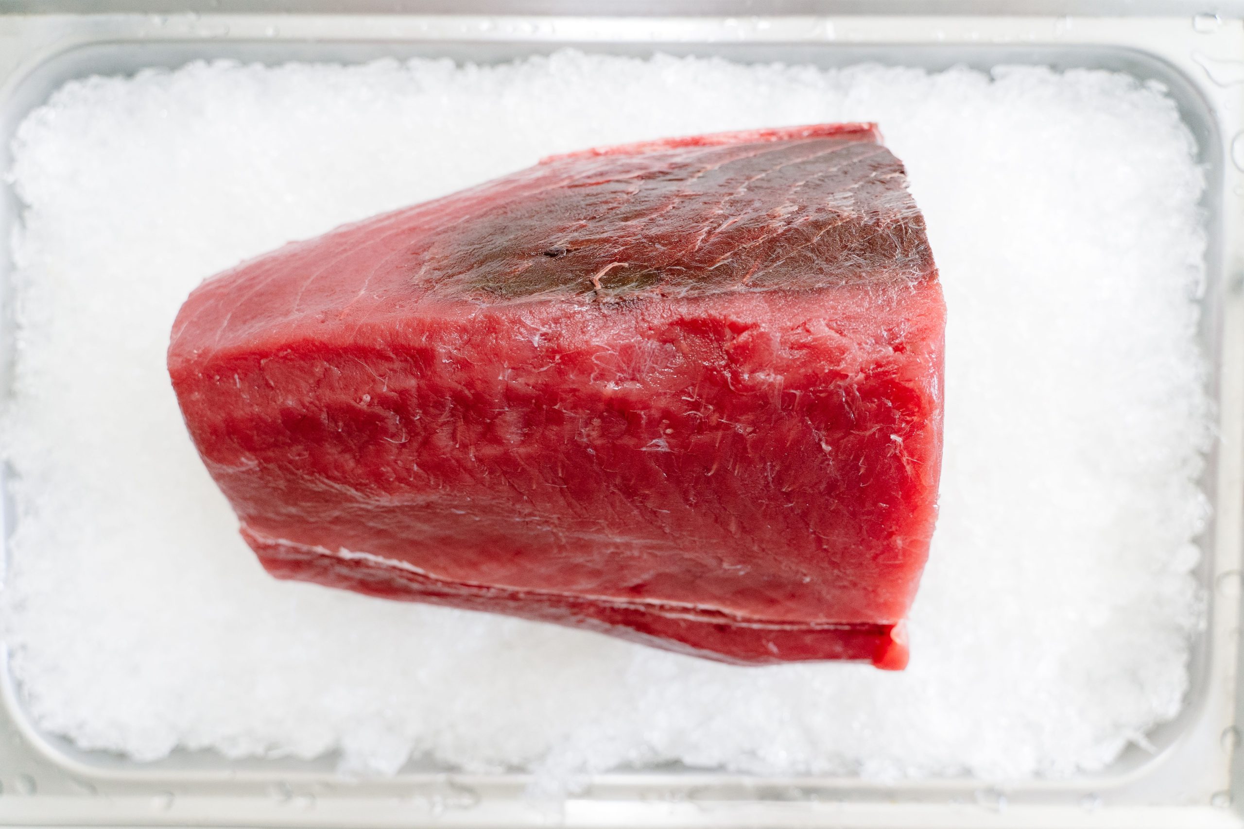
Who would have thought that tuna fish bones, which are usually discarded as waste at fish processing sites, could become a candidate material for protecting dental pulp? This was demonstrated by Prof. drg. Tetiana Haniastuti, M.Kes., Ph.D., and her team from the Department of Oral Biology, Faculty of Dentistry, Universitas Gadjah Mada. Their study, published in the International Journal of Dentistry in November 2020, showed that hydroxyapatite synthesized from yellowfin tuna bones (Thunnus albacares) is not only safe for human dental pulp cells, but also promotes their growth and differentiation.
From Fish Bones to Biomaterials
Yellowfin tuna is one of the most widely consumed fish species in Indonesia. Its meat ends up on dinner tables, but its bones? They almost always end up as waste. Yet tuna bones contain high levels of calcium and phosphorus—two elements that can be processed into hydroxyapatite (HA), a mineral compound whose chemical composition closely resembles that of hard tissues in the human body, such as bones and teeth.
In this study, the tuna bones were cleaned, pressure-heated, dried for four days, and then sintered at 700°C before being ground into powder. This powder was then tested on human dental pulp cells isolated from the impacted third molars of healthy patients aged 18 to 24 years.
Why dental pulp? Because the pulp is living tissue located at the heart of the tooth, making it vulnerable to injury from restorative procedures, caries removal, and trauma. When the pulp is injured, a pulp capping material is needed to stimulate reparative dentin formation while maintaining tissue vitality. Calcium hydroxide has long been considered the gold standard for this purpose. However, this material also has limitations: its highly alkaline pH can irritate pulp tissue and even trigger necrosis in the underlying cell layer.
Cells Remained Viable—and Even Increased
The research team tested pulp cell viability using the MTT assay, a standard method for measuring the mitochondrial activity of living cells. The cells were exposed to various concentrations of THA (tuna-derived hydroxyapatite) powder, ranging from 6.25 to 200 µg/ml, for 24, 48, and 72 hours.
The results were well above the safety threshold. Cell viability ranged from 91 to 116 percent across all observation periods. According to ISO 10993-5, a material is considered non-cytotoxic when cell viability is above 80 percent. THA not only exceeded this threshold, but at the highest concentration (200 µg/ml) after 72 hours, the number of viable cells actually exceeded that of the untreated control group.
Microscopic examination using ethidium bromide-acridine orange staining revealed elongated spindle-shaped cells with oval nuclei, forming linear arrangements resembling bundles—a morphology characteristic of healthy dental pulp cells that are actively proliferating. No suspicious nuclear changes or apoptotic bodies were observed.
A Key Enzyme Was Also Stimulated
The most interesting part of the study was the finding regarding alkaline phosphatase (ALP) activity. This enzyme is an early marker of odontogenic differentiation, the process by which pulp cells begin to transform into odontoblasts capable of forming dentin matrix.
The pulp cells were cultured in odontoblastic differentiation medium and exposed to THA for 7, 11, and 15 days. The results were consistent: the higher the THA concentration, the higher the ALP activity. Groups treated with 100 and 200 µg/ml THA showed significantly higher ALP activity than the control group at all observation periods (p < 0.05).
“The ALP activity test showed that THA increased alkaline phosphatase activity in pulp cells, suggesting its potential to promote dentin matrix formation, which is important for pulp repair following injury.”
This finding is consistent with previous studies showing that hydroxyapatite-based scaffolds can induce the proliferation and osteogenic differentiation of human dental pulp stem cells.
A Long Road Toward Clinical Application
Prof. Tetiana and her team emphasized that this study was only an in vitro study, meaning that it was conducted in a laboratory setting rather than on actual patient teeth. Many stages remain before THA can be used clinically: animal studies to evaluate dentin bridge formation and inflammatory responses, development of an optimal physicochemical formulation, and eventually clinical trials in humans.
Nevertheless, the potential demonstrated by the study is sufficient to make the research strategically relevant. Indonesia is one of the world's largest tuna producers. Utilizing abundant fish-bone waste as a source of dental biomaterials is not only a matter of scientific innovation, but also of resource efficiency and sustainability.
The question that remains is: how far can these tuna fish bones go—from the laboratory bench to the dental chair?
Source DOI: https://doi.org/10.1155/2020/8857534
Authors: Anny Anggraini; drg. Achmad Zam Zam Aghasy, M.Kes.
Photo: Pexels