
Imagine a 10-year-old child born with a cleft in the lip and palate. After years of surgeries and treatment, the child arrives at an orthodontic clinic with one lingering question: can their teeth be aligned like those of other children? The question may seem simple, but it reflects biological complexities that have received relatively little scientific attention.
A systematic review published in The Cleft Palate Craniofacial Journal sought to answer this question. A research team from the Faculty of Dentistry, Universitas Gadjah Mada (UGM), including lead researcher drg. Ananto Ali Alhasyimi, DDS, MDSc, M.Clin.Dent(orth), PhD, together with drg. Anrizandy Narwidina, DDS, MDSc, Sp.KGA, PhD, from the Department of Pediatric Dentistry, screened 528 research records from around the world to address a clinical question that has long been overlooked: what actually happens to patients' teeth after secondary alveolar bone grafting?
Filling the Gap, Opening the Path to Eruption
Cleft lip and palate (CLP) is one of the most common congenital anomalies. Its treatment is not simply about anatomically closing the cleft, but also about restoring function, facial aesthetics, and long-term occlusal stability. This is where secondary alveolar bone grafting (SABG) becomes crucial. The procedure involves placing bone, generally harvested from the iliac crest, into the alveolar cleft to restore continuity of the maxillary arch.
What makes this review particularly interesting is that it goes beyond confirming the surgical success of SABG. The researchers shifted their focus from the operating room to the orthodontic chair. Of the 528 research records screened, only six studies met the strict inclusion criteria, involving a total of 167 patients. The results were encouraging: in patients whose grafts integrated successfully, orthodontic tooth movement into the grafted bone was feasible. No graft failures were reported as a direct consequence of orthodontic mechanics.
Findings concerning maxillary canine eruption were among the most clinically meaningful. The permanent canine is the tooth most frequently affected in patients with CLP, and its eruption through the cleft region serves as an indicator of successful rehabilitation. Of the four studies involving 112 patients that evaluated this outcome, most reported successful spontaneous or orthodontically guided eruption, particularly when SABG was performed before the permanent canine began to erupt, during the mixed dentition stage.
When CT Scans Reveal Bone and Periodontal Tissue
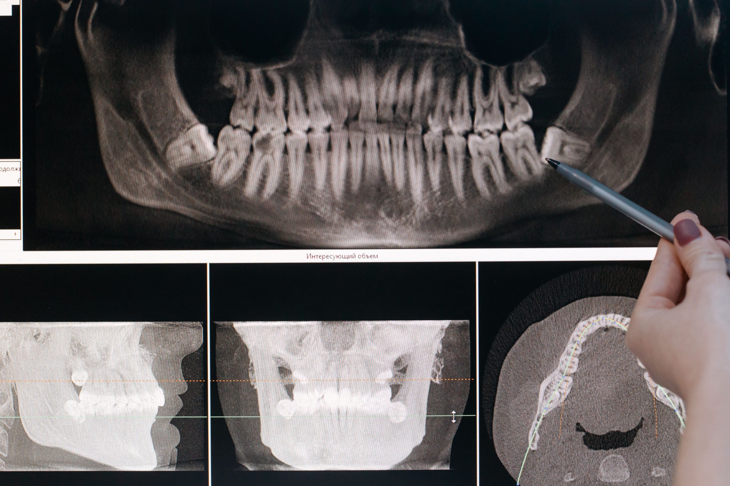
Evaluation of periodontal health and alveolar bone morphology following orthodontic treatment provides a more nuanced picture. Using cone-beam computed tomography (CBCT), one cross-sectional study involving 30 patients found that moving the canine into the grafted region produced generally acceptable alveolar bone morphology. Buccal and lingual bone plate thickness was maintained, alveolar crest height was preserved, and no clinically significant bone dehiscence was identified.
However, there was an important caveat. The buccal bone plate on the cleft side tended to be thinner than that on the healthy side. This does not indicate treatment failure, but it serves as a reminder that reconstructed tissue is not identical to the original tissue and that long-term monitoring remains necessary.
“Current evidence indicates that SABG establishes a clinically viable foundation for subsequent orthodontic rehabilitation in patients with cleft lip and palate. Following successful osseous integration, both directed orthodontic tooth movement and the eruption of maxillary canines into the grafted site are highly feasible.”
This was the conclusion reached by the research team in the article—a statement that is cautious yet meaningful for clinicians who work with CLP patients every day.
Low Certainty, High Relevance
The systematic review did not stop at optimism. Using the GRADE framework to assess the quality of evidence, the researchers honestly stated that the evidence for orthodontic feasibility and canine eruption was of low certainty, while the evidence for periodontal outcomes was of very low certainty. This was primarily due to the predominance of observational study designs, heterogeneity in orthodontic protocols across studies, and small sample sizes.
This does not diminish the findings. Rather, it reflects scientific transparency that strengthens the value of the review. In a complex clinical setting such as CLP management, where every decision affects a child's physical and psychological development, distinguishing between “this appears clinically successful” and “this has been definitively proven” is crucial.
The researchers' main recommendation is for prospective, multicenter clinical trials with standardized orthodontic protocols, longer follow-up periods, and integrated assessments of surgical, orthodontic, periodontal, and patient-reported quality-of-life outcomes within a coherent framework. Such studies, if conducted, could fill the current evidence gap that leaves clinicians relying on a combination of available evidence and individual clinical expertise.
For that 10-year-old child sitting in the orthodontic chair, and for thousands of other CLP patients waiting for certainty about their smiles, the scientific journey is not yet over. But at least, the direction is now a little clearer.
Source DOI: http://10.1177/10556656261454065
Authors: Nanda Ayu, drg. Achmad Zam Zam Aghasy, M.Kes.
Photo: Pexels