
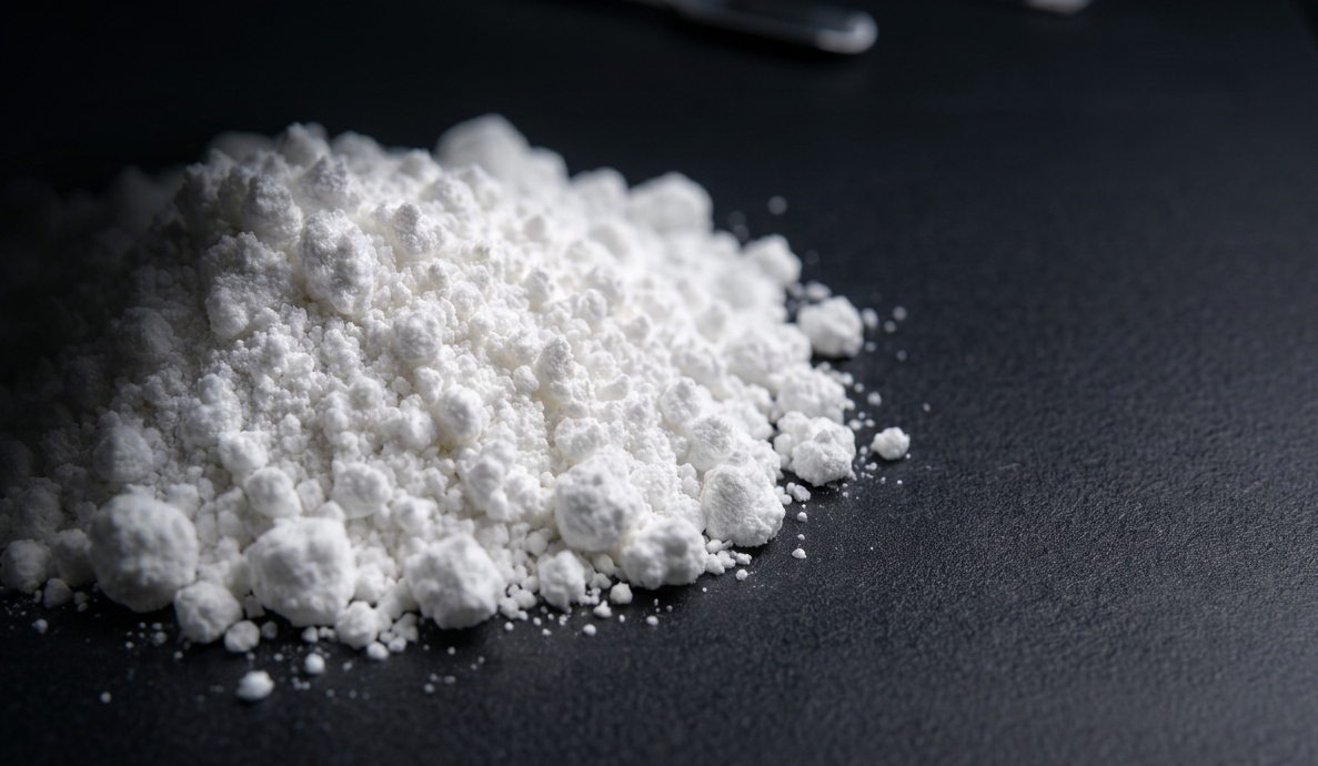
Calcium hydroxide paste, which has long been relied upon by dentists to eliminate bacteria inside root canals, may carry a hidden risk that has received limited clinical attention: the longer the paste remains inside the root canal, the more fragile the remaining tooth structure becomes. This is the key finding of an in vitro study conducted by Prof. drg. Diatri Nari Ratih, M.Kes., Ph.D., Sp.KG(K), from the Department of Conservative Dentistry, Faculty of Dentistry, Universitas Gadjah Mada, Yogyakarta. The study, published in The Indonesian Journal of Dental Research, used 72 extracted human mandibular premolars. The results were striking: exposure to calcium hydroxide for 14 to 30 days significantly reduced the flexural strength of root canal dentin compared with the control group.
Calcium hydroxide, or Ca(OH)₂, is not a new material. For decades, it has been a mainstay in endodontic treatment, particularly as an intracanal medicament—a material placed inside the root canal between treatment visits. Its mechanism is simple yet effective: the hydroxyl ions released by this material create a highly alkaline environment that eliminates bacteria while simultaneously stimulating mineralised tissue formation.
However, the same chemical properties that make calcium hydroxide effective against bacteria may also become a concern. Dentin, the hard tissue layer inside the tooth root, consists of approximately 22% organic material, most of which is type I collagen. This collagen provides dentin with its ability to withstand stress and bending forces. When calcium hydroxide remains in prolonged contact with dentin, hydroxyl ions that diffuse through the dentinal tubules are suspected of damaging this organic matrix by disrupting the interaction between collagen fibrils and mineral crystals that form the dentin structure.
The question addressed by Diatri Nari Ratih’s research was simple yet clinically significant: how long can calcium hydroxide remain inside the root canal without causing meaningful structural damage to the tooth? To answer this question, 72 extracted single-rooted mandibular premolars free from caries and cracks were used as samples. Each tooth was sectioned to obtain an 18-mm-long root, after which the root canals were cleaned and shaped using the crown-down technique with ProTaper instruments.
Following preparation, the teeth were divided into three groups. The first group received calcium hydroxide mixed with sterile saline solution. The second group received a commercial calcium hydroxide product, UltraCal®. The third group served as the control group and was filled only with saline solution. All teeth were sealed at the apical and coronal regions using composite resin, immersed in artificial saliva, and stored in an incubator at 37°C to approximate human oral conditions.
On days 7, 14, and 30, eight teeth from each group were removed. The internal root canal dentin was sectioned into small bars measuring 1 × 1 mm with a length of 7 mm. These dentin specimens were then tested using a three-point bending test with an MTS universal testing machine to measure their flexural strength, or the amount of force required to fracture them.
The results were clear. On day 7, differences in flexural strength among the three groups were not statistically significant. However, by days 14 and 30, calcium hydroxide—both the saline-mixed preparation and the commercial UltraCal® product—had significantly reduced dentin flexural strength compared with the control group.
The numerical differences were substantial. Normal dentin flexural strength was approximately 212.9 MPa. In the group exposed to calcium hydroxide mixed with saline for 30 days, flexural strength decreased to approximately 145.72 MPa. This represents a reduction of nearly 32% within only one month. The UltraCal® group showed a more moderate decline, reaching approximately 156.42 MPa after 30 days, but this value remained considerably lower than normal dentin strength.
“Exposure to calcium hydroxide for 14 and 30 days can significantly reduce the flexural strength of root canal dentin. The longer the exposure period, the greater the effect.”
This was the conclusion drawn from the study data. Two-way ANOVA followed by Tukey’s post-hoc test confirmed that differences between groups and immersion periods were statistically significant.
Another interesting finding was that calcium hydroxide mixed with saline appeared to cause greater damage compared with the commercial UltraCal® product. This difference is associated with surface tension. Calcium hydroxide mixed with saline has lower surface tension, allowing deeper penetration into dentinal tubules. As a result, its destructive effect on the dentin collagen matrix may become more pronounced.
This finding is important for clinical practice. Many dentists still prepare calcium hydroxide by mixing it with saline because it is more economical. Although this approach may provide comparable antimicrobial effects, the study suggests that it carries a higher risk of structural damage to root dentin.
The mechanism of damage is thought to occur through two pathways. First, the high pH caused by hydroxyl ions weakens the organic matrix of dentin, including collagen degradation through denaturation and hydrolysis processes. Second, calcium hydroxide facilitates tissue detachment from dentin walls, making the dentin surface more vulnerable to crack initiation and propagation.
The clinical implications of this study are clear. Seven days of calcium hydroxide exposure did not produce significant differences in dentin flexural strength. This finding corresponds with common clinical practice, where dentists typically place intracanal medicaments for approximately one week before the next appointment.
However, when the interval between visits becomes longer—whether due to logistical issues, patient conditions, or clinical decisions—the risk to dentin structural integrity increases significantly. Teeth that have undergone root canal treatment have already lost part of their structure due to canal preparation and access cavity formation. Weakening of dentin caused by prolonged calcium hydroxide exposure may further increase susceptibility to root fractures.
This study recommends that calcium hydroxide use as an intracanal medicament should not exceed seven days. This does not mean that the material should be avoided. On the contrary, calcium hydroxide remains an effective and trusted medicament when used within an appropriate time frame.
The study also highlights several areas requiring further investigation. One of them is the combined effect of calcium hydroxide and sodium hypochlorite (NaOCl), an irrigation solution increasingly used alongside calcium hydroxide. Both materials are known to have synergistic effects in dissolving tissue, but the combined impact on dentin mechanical strength has not yet been fully understood.
Another question concerns the choice of control material. This study used saline as the control; however, the findings indicated that saline itself slightly reduced dentin flexural strength, possibly due to the dissolving effects of chloride ions. Future studies may consider distilled water as a more neutral control.
For patients, studies like this serve as a reminder that root canal treatment is not merely about cleaning the tooth. It is a complex procedure involving biomechanical considerations. Teeth treated carefully—including appropriate management of intracanal medicament duration—are likely to have better long-term outcomes.
Author: drg. Achmad Zam Zam Aghasy, M.Kes., Annisa Dwi Noviyanti
Photo: FreePik