
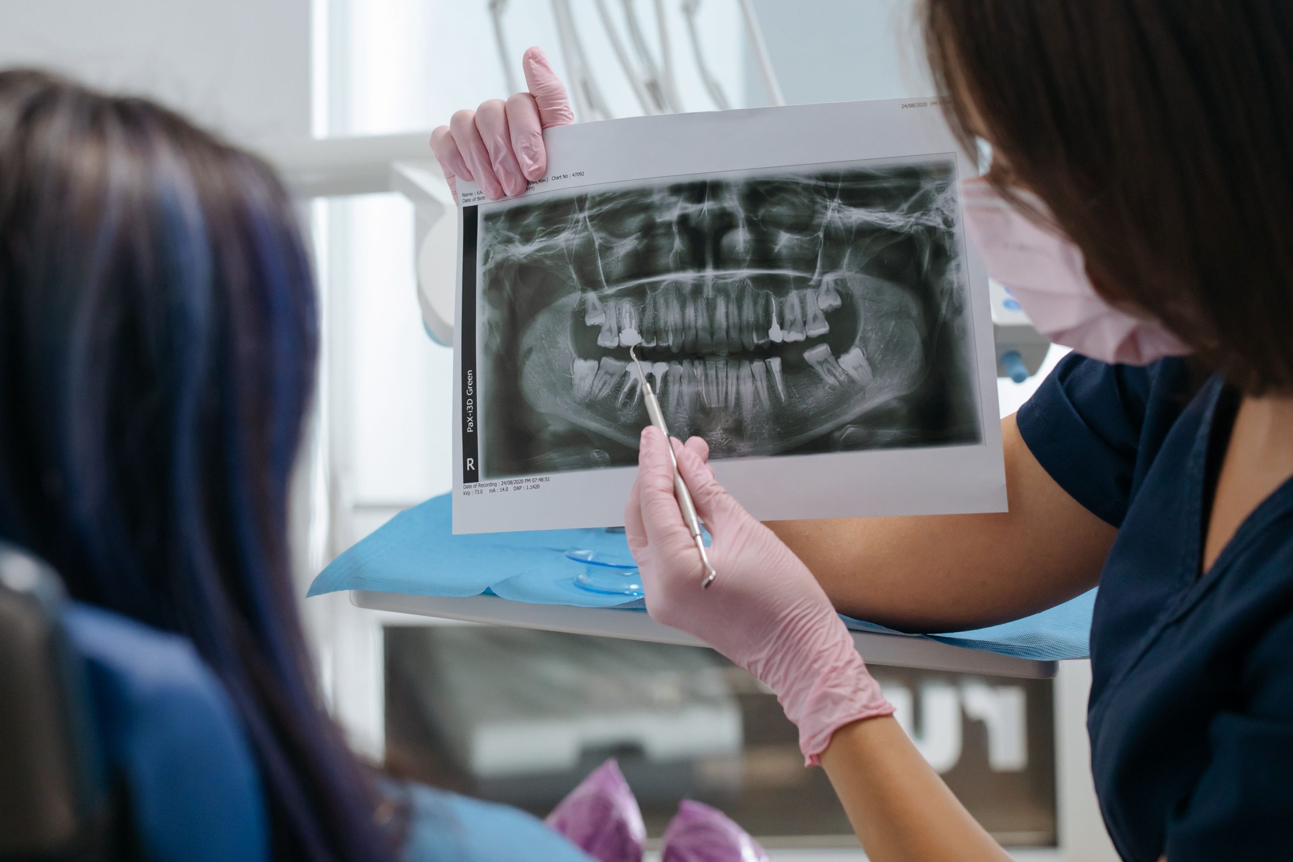
Ten days after a panoramic X-ray examination, something invisible occurs inside the patient’s mouth. The composition of epithelial cells in the buccal and gingival mucosa shifts: superficial cells decrease, while intermediate cells increase significantly.
These changes are not clinical symptoms that patients can feel, but they can be clearly observed under a light microscope at 400× magnification. This was the main finding of a study published in the Brazilian Journal of Oral Sciences by researchers from the Faculty of Dentistry, Universitas Gadjah Mada, led by drg. Ryna Dwi Yanuaryska, Ph.D. The study was the first to specifically evaluate oral epithelial maturation patterns following exposure to three commonly used dental radiographic methods: analog panoramic radiography, digital panoramic radiography, and cone beam computed tomography (CBCT).
Cells Shift Without Causing Symptoms
The oral mucosa functions like a regeneration machine that never stops. New cells originate in the parabasal layer, migrate upward, mature into intermediate cells, and eventually become superficial cells on the outermost surface before naturally being shed. This process is known as normal epithelial maturation, and disruption of this process is considered an early marker of potentially pathological cellular changes.
Researchers have previously demonstrated that X-ray exposure can cause cell death through cytotoxic mechanisms, characterized by pyknosis, karyolysis, and karyorrhexis. But does exposure also disrupt the overall process of cellular maturation? That was the question that prompted drg. Ryna and her team to design this study.
A total of 40 subjects aged 25 years and older who met strict eligibility criteria were included. They were divided into four groups: three radiographic-exposure groups and one unexposed control group. All participants were non-smokers, did not consume alcohol, and had no visible mucosal lesions. Cytological smears were collected from the buccal and gingival mucosa before exposure and again ten days afterward. Papanicolaou staining was used to determine the proportions of parabasal, intermediate, and superficial cells.
Digital Panoramic Radiography Shows the Most Pronounced Effect
The results were quite striking. In the control group, there were no significant changes in cell composition during the ten-day observation period. In contrast, all three radiographic methods produced similar changes: intermediate cells increased significantly (p < 0.05), while superficial cells decreased significantly (p < 0.05) in both the buccal and gingival mucosa.
Interestingly, digital panoramic radiography produced the largest shift among the three methods. In the buccal mucosa, the proportion of intermediate cells increased from an average of 69.70% to 81.20% after digital panoramic exposure, while superficial cells declined sharply from 30.50% to 17.83%. These changes were greater than those observed in the CBCT and analog panoramic groups.
“This study is the first to assess maturation patterns following dental imaging using a cytopathological method. Our results show that exposure to analog/digital panoramic radiography and CBCT can induce cytotoxicity by altering the maturation patterns of buccal and gingival mucosal cells.” — drg. Ryna Dwi Yanuaryska, Ph.D., Faculty of Dentistry, UGM
There was no significant difference between the buccal and gingival mucosa in terms of the magnitude of cellular changes, suggesting that the two tissues respond to radiation in broadly similar ways.
This Does Not Mean Dental X-Rays Should Be Avoided
These findings are not a call to stop using dental radiography. Panoramic radiography and CBCT remain essential diagnostic tools in dental care, from evaluating impacted teeth and planning implants to detecting jaw abnormalities that cannot be identified clinically. Without radiographic imaging, many diagnoses could be missed.
However, the study provides strong scientific support for a principle that has long been recommended in radiation protection: the ALARA principle (as low as reasonably achievable). This means that radiation exposure should be kept to the minimum necessary and that every X-ray examination should be based on a clear clinical indication rather than routine or habit.
The shift in cellular maturation patterns observed in this study resembles changes that occur in mucosal tissue following exposure to tobacco and alcohol, two established risk factors for oral cancer. This does not mean that dental X-rays are equivalent to smoking. Rather, the findings serve as a reminder that ionizing radiation, at any level, does not act without biological consequences.
The researchers also recommend further studies measuring the keratinization index, namely the proportion of anucleated superficial cells, as an additional marker for the early detection of cellular changes associated with oral cancer. This represents an important area for further investigation.
For patients wondering whether their annual dental X-rays are safe, the answer remains yes, when clinically necessary. But “necessary” is the key word that should never be overlooked.
Source DOI: doi: 10.1016/j.mrgentox.2004.05.008.
Authors: Anny Anggraini; drg. Achmad Zam Zam Aghasy, M.Kes.
Photo: Pexels