
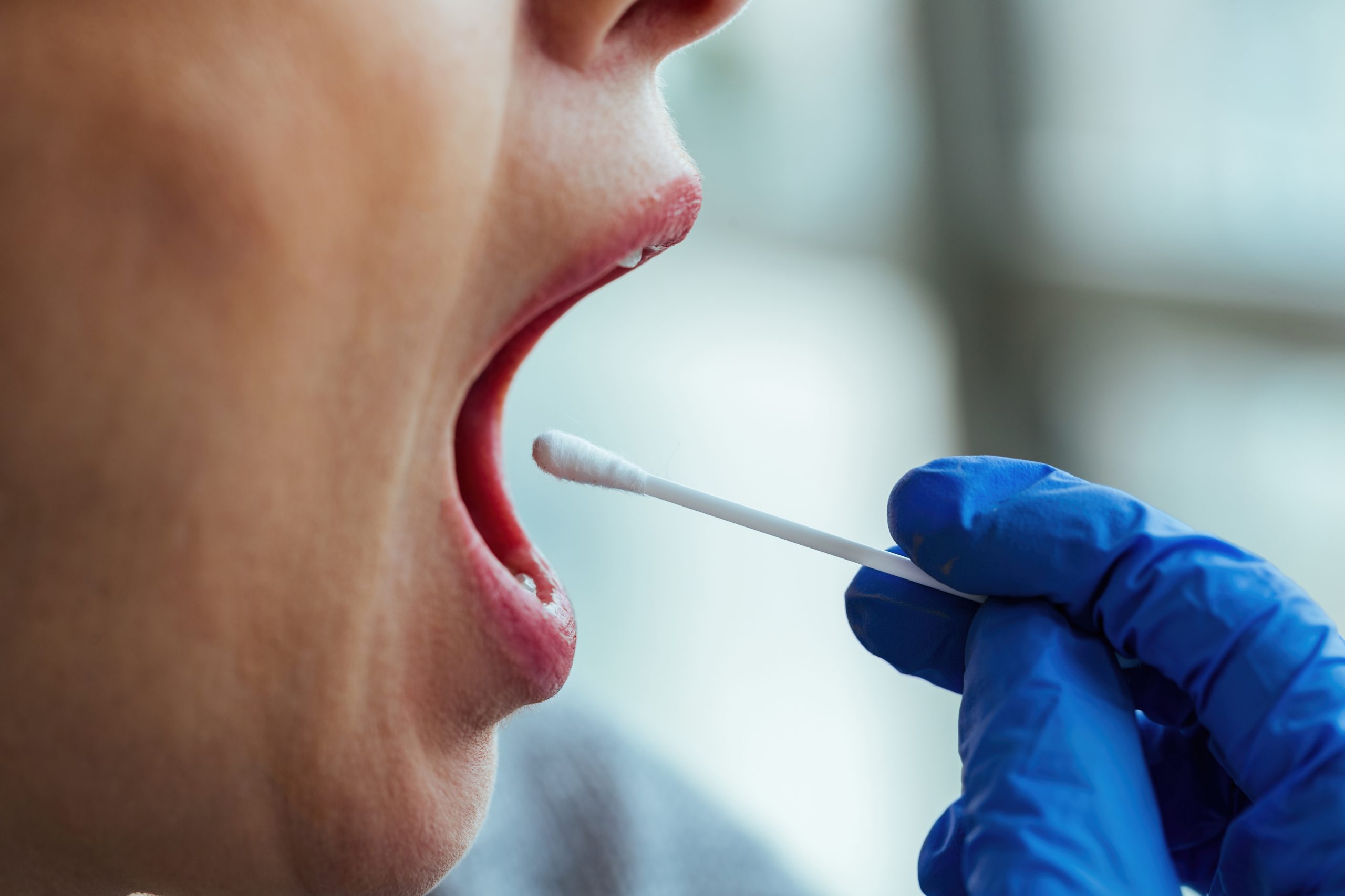
Imagine a tumor growing aggressively at the base of the tongue, spreading to the lymph nodes in the neck, and recurring even after radical surgery. This is the reality of base-of-tongue cancer, which has been an oncologist's nightmare for decades. Patient survival rates have barely improved, while chemotherapy and radiotherapy have inherent limitations. This is where Prof. drg. Supriatno, M.Kes., MDSc., PhD., from the Department of Oral Medicine, Faculty of Dentistry, Universitas Gadjah Mada, has attempted to open a different path: injecting a tumor-suppressor gene directly into cancerous tissue using electrical pulses as a guide.
When Electricity Opens the Cellular Door
The technique used in this study is known as electro-gene therapy, or electroporation. The concept is relatively simple, but its implementation requires considerable precision: electrical pulses of a specific intensity are delivered to tumor tissue, temporarily “opening” the membranes of cancer cells for long enough to allow foreign DNA injected from outside to enter.
In this experiment, Prof. Supriatno, together with drg. Inne Suherna Sasmita from the Faculty of Dentistry, Universitas Padjadjaran, used the plasmid pcDNA3.1-p27^Kip1 wild type (wt) as the genetic “payload.” The p27^Kip1 gene is a cyclin-dependent kinase inhibitor that suppresses the cell cycle during the G1 phase. In many types of human cancer, expression of this gene is dramatically reduced, allowing tumor cells to proliferate without restraint.
The model used was a Balb/c nude mouse xenograft with SP-C3 cancer cells isolated from cervical lymph-node metastasis in a patient with base-of-tongue cancer. These cells are far from ordinary: SP-C3 is known for its extremely rapid growth rate, high invasive capacity, and rapid local recurrence following radical excision.
Gene Enters, Tumor Shrinks
The results were striking. Gene transfection efficiency using a combination of electroporation and plasmid injection reached 55–70% of cells, far exceeding that achieved through the injection of “naked” DNA without electrical pulses, which resulted in only a small number of positive cells. This was confirmed through expression of the reporter gene enhanced green fluorescent protein (EGFP), which was detected under fluorescence microscopy.
More importantly, tumors in the group receiving pcDNA3.1-p27^Kip1 wt grew significantly more slowly than those in the control group, which received only an empty vector. Western blotting analysis confirmed that p27^Kip1 protein expression was significantly increased in the transfected tumor tissue.
“In vivo gene transfer method is a simple procedure and can solve some of the critical drawbacks of the present gene transfer techniques, thus providing a new strategy for gene therapy.”
This statement appears in the paper's conclusion and reflects measured optimism: it is not a claim of a cure, but rather the opening of a new strategy worthy of further investigation.
Apoptosis Activated, Not Merely Growth Inhibited
One of the most interesting findings of this study is evidence that the p27^Kip1 gene does more than inhibit tumor growth; it also triggers programmed cancer cell death.
Proteolytic activity of caspase-3 in transfected tumors increased 2.9-fold compared with controls, while caspase-9 activity increased 1.7-fold, with both showing statistical significance at P = 0.001. Caspase-3 and caspase-9 are key enzymes in the intrinsic apoptotic pathway. This means that the p27^Kip1 gene does not merely “freeze” cancer cells in the G1 phase; it also drives those cells toward orderly cell death.
Notably, no weight loss in the mice or burns on the skin around the electrode sites were observed during the experimental period. This indicates that electroporation using a voltage of 80 V for a xenograft measuring 1.0 cm in diameter was relatively safe locally.
The Long Road to the Clinic
There are, of course, limitations that must be understood. Prof. Supriatno himself noted that this therapeutic approach remains limited to localized tumors. Multiple metastases located far from the electrodes cannot yet be efficiently reached using this system. In the future, the researchers plan to combine p27^Kip1 gene transfer with other genes and anticancer agents to expand its therapeutic reach.
Nevertheless, in the context of oral cancer, whose mortality-to-incidence ratio has remained nearly stagnant at 0.47–0.48 for two decades, every new strategy demonstrating preclinical efficacy deserves serious attention. Electro-gene therapy using p27^Kip1 offers something that conventional approaches do not: the ability to insert genetic instructions directly into a tumor, forcing cancer cells to stop growing from within and ultimately causing them to destroy themselves.
Base-of-tongue cancer may be one of the most stubborn forms of oral cancer. But genes, it seems, can speak a language that the cells themselves understand.
Source DOI: https://doi.org/10.24198/pjd.vol21no3.14106
Authors: Anny Anggraini; drg. Achmad Zam Zam Aghasy, M.Kes.
Photo: Freepik