
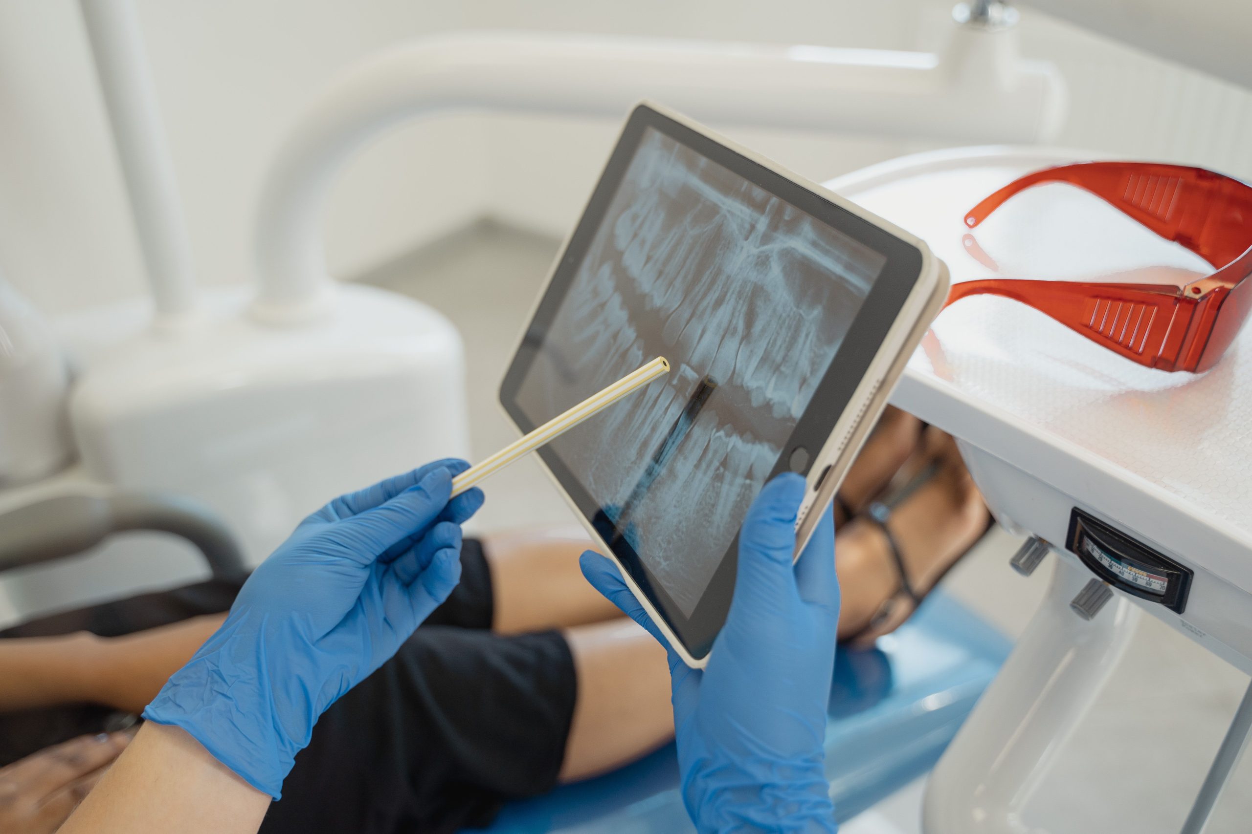
Imagine a dentist who has to examine hundreds of panoramic radiographs every day, assess the condition of the alveolar bone around each tooth one by one, and then determine whether a patient has mild, moderate, or severe periodontitis. The work is exhausting, and fatigue creates opportunities for errors. This prompted Dr. drg. Rini Widyaningrum, M.Biotech, from the Department of Dentomaxillofacial Radiology, Faculty of Dentistry, Universitas Gadjah Mada (UGM), to ask a seemingly simple question: could a computer do it?
The answer, it turns out, is yes—and the results exceeded expectations.
When Radiographs Meet Deep Learning
Periodontitis is a chronic inflammatory disease that progressively damages the tissues supporting the teeth. If left untreated, alveolar bone loss can lead to tooth mobility and tooth loss, ultimately impairing chewing and swallowing and reducing overall quality of life. Accurate diagnosis is therefore essential, and panoramic radiography is one of the primary diagnostic tools—a two-dimensional radiographic image that captures the entire dental arch at once.
Published in Imaging Science in Dentistry in 2022, this study compared two deep learning models for image segmentation on panoramic radiographs: Multi-Label U-Net and Mask R-CNN. The objective was to automatically detect and stage periodontitis based on the degree of radiographic bone loss (RBL).
The research team used 100 digital panoramic radiographs from patients at the UGM Dental Hospital, acquired between May and June 2017. The images were manually annotated by dentists and periodontists, who established a consensus classification based on the latest system from the 2017 World Workshop on the Classification of Periodontal and Peri-Implant Diseases. Data augmentation increased the original 100 images to 1,100 images, containing a total of 9,907 regions of interest (ROIs) divided into five classes: normal and stages 1 through 4.
Two Models, Two Approaches, One Winner
U-Net operates on the principle of semantic segmentation—classifying each pixel in an image into a particular category to produce a comprehensive map of bone conditions. Its architecture resembles the letter “U”: the left side compresses information through the encoder, while the right side reconstructs it through the decoder, with direct connections between the two sides to preserve spatial details.
Mask R-CNN works differently. It performs instance segmentation, allowing it to distinguish individual objects—in this case, the bone surrounding each tooth separately. This approach is theoretically more detailed but also more complex.
The results were striking. Multi-Label U-Net achieved a Dice coefficient of 0.96 and an IoU score of 0.97, figures approaching the upper end of performance standards for medical image segmentation. Mask R-CNN lagged behind, with a Dice coefficient of 0.87 and an IoU score of 0.74. Nevertheless, Mask R-CNN still achieved a detection accuracy of 95%, with 85.6% precision, 88.2% recall, and an F1-score of 86.6%.
“Multi-Label U-Net produced superior image segmentation to that of Mask R-CNN. The authors recommend integrating it with other techniques to develop hybrid models for automatic periodontitis detection.” — Dr. drg. Rini Widyaningrum, M.Biotech, et al., Imaging Science in Dentistry, 2022
Interestingly, Mask R-CNN performed best in detecting stage 4 periodontitis—the most severe condition, characterized by the loss of five or more teeth—with a precision of 0.97 and recall of 0.95. This suggests that each model has strengths that could complement those of the other.
From Manual Annotation to Intelligent Automation
One of the greatest challenges in this study was not the algorithm itself, but the data. The imbalanced distribution of samples—where normal and stage 1 cases were much more numerous than stage 4 cases—could cause the model to learn with bias. The researchers addressed this through data augmentation by rotating images between −5° and 5°, generating sufficient variation to train the model without underfitting.
The annotation process itself was conducted with great precision. Each alveolar bone crest and wall surrounding every tooth was marked with a square box on the panoramic radiograph, accompanied by a stage code based on the percentage of RBL. Bone surrounding severely crowded teeth was deliberately left unannotated to avoid ambiguity.
As a result, the trained model did more than simply recognize general patterns; it learned the nuances of dental anatomy within a genuine clinical context.
A Tireless Machine, More Focused Dentists
This research is not about replacing dentists or radiologists. It is about giving them a more powerful tool.
Fatigue and excessive workloads are real factors that can affect the accuracy of radiographic diagnosis. A deep learning-based system capable of pre-screening and providing preliminary labels on panoramic radiographs could allow dentists to devote more attention to complex clinical decisions rather than repetitive measurement tasks.
Dr. drg. Rini Widyaningrum and her team recommend developing a hybrid model that combines the strength of Multi-Label U-Net in high-precision segmentation with the ability of Mask R-CNN to distinguish individual objects. If successfully implemented, such a combination could serve as the foundation for an automated periodontitis diagnostic system that is genuinely ready for clinical use.
The question is no longer whether machines can read radiographs. The question now is: how quickly can we build a system intelligent enough to help dentists make the best decisions for their patients?
Source DOI: 10.5624/isd.20220105
Authors: Anny Anggraini; drg. Achmad Zam Zam Aghasy, M.Kes.
Photo: Pexels