
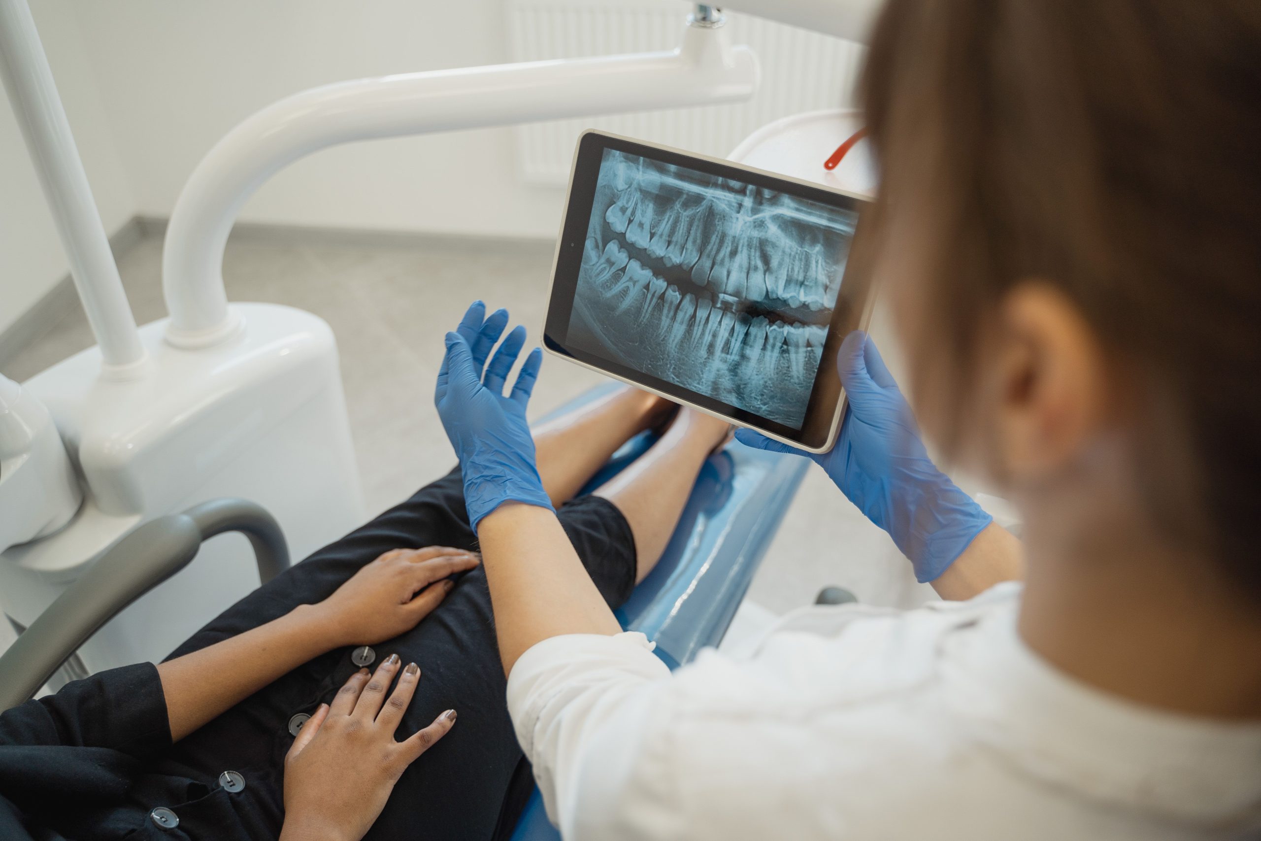
Until now, dental X-rays have largely been regarded as the domain of dentists: examining caries, assessing root positions, or detecting infections around the root apex. But Dr. drg. Rini Widyaningrum, M.Biotech., from the Department of Dentomaxillofacial Radiology, Faculty of Dentistry, Universitas Gadjah Mada, had a different perspective. She believed that the trabecular bone patterns visible in periapical radiographs contained much more information—including signs of osteoporosis, which until now could only be confirmed using expensive and not always readily accessible DEXA equipment.
That belief now has scientific evidence behind it. A study published in the International Journal of Dentistry in February 2023 showed that an automated machine-learning-based segmentation method could detect osteoporosis from periapical radiographs with an accuracy of 90.48%.
Bones That Tell a Story Through Pixels
Osteoporosis is not simply a matter of fragile bones. The condition increases the risk of fractures, exacerbates periodontal tissue damage, and may contribute to tooth loss and jawbone erosion. Ironically, early osteoporosis detection still relies on DEXA scanning (dual-energy X-ray absorptiometry), a procedure that requires specialized equipment, can be costly, and is not available at every healthcare facility.
This is where dental radiographs offer a promising alternative. Changes in trabecular bone—the fine, mesh-like network within the jawbone—can actually be observed on periapical radiographs routinely taken in dental clinics. The problem is that manual interpretation depends on the clinician's experience and is vulnerable to fatigue. Interpretations may also vary between clinicians.
The research team led by Dr. drg. Rini Widyaningrum proposed a solution: let computers read the bone instead.
How the Machine Learned to Read Bone
The study used 102 periapical radiographs from the anterior and posterior regions of the mandible, retrospectively collected from postmenopausal Javanese women aged 58–81 years. The data were obtained from the UGM Dental Hospital, with osteoporosis diagnoses confirmed by DEXA scans at Dr. Sardjito General Hospital, Yogyakarta. Of the total dataset, 60 images were used for training and 42 for testing.
The proposed method consisted of five stages. First, MATLAB-based software identified the region of interest (ROI) in the trabecular area, at least 2 mm from the tooth apex. A 300 × 400-pixel area was then automatically cropped from that point. The ROI was converted to grayscale, segmented using a color-clustering algorithm, and its pixel distribution was extracted before classification using three machine-learning methods: decision tree, naive Bayes, and multilayer perceptron.
The researchers compared two segmentation methods: K-means and Fuzzy C-means. The results were clear: K-means performed better. K-means segmentation produced better image contrast and achieved an overall training performance of 81.67%, compared with only 75.28% for Fuzzy C-means.
“Our proposed method makes a valuable contribution to medical image analysis for osteoporosis detection and can be further developed for applications in dental practice.” — Dr. drg. Rini Widyaningrum, M.Biotech., Department of Dentomaxillofacial Radiology, Faculty of Dentistry, UGM
The Best Combination: K-Means and Multilayer Perceptron
Of the six algorithm combinations tested, one emerged as the strongest: K-means with 10 clusters combined with a multilayer perceptron as the final classifier. On the test dataset, this combination achieved 90.48% accuracy, 90.90% specificity, and 90.00% sensitivity.
In other words, of the 42 test images, only four were incorrectly classified: two osteoporosis cases were classified as normal, while two normal cases were classified as osteoporosis. For a method based on routine dental radiographs, these figures are remarkably high.
Equally important, the detection process took less than one minute, measured from the point at which the ROI was extracted from the radiograph. This speed could be particularly valuable in healthcare facilities handling large numbers of patients within limited time.
Dental X-Rays as a Gateway to Early Detection
The study addresses an issue that has received relatively little attention: dentists could become frontline professionals in osteoporosis detection, rather than leaving this responsibility solely to bone specialists or medical radiologists. In developing countries such as Indonesia, where access to DEXA scans remains limited, periapical radiographs already routinely obtained in dental clinics could provide a more affordable and equitable entry point for screening.
There are, of course, limitations. The sample size was relatively small, and all data came from postmenopausal Javanese women. Therefore, broader generalization will require further studies involving larger and more diverse populations.
Nevertheless, the direction is clear. As artificial intelligence becomes increasingly integrated into clinical practice, a seemingly simple dental X-ray may contain far more information than previously imagined. Without requiring any additional equipment, dentists may already be holding a key to the early detection of a disease affecting the bones of millions of Indonesians.
Source DOI: https://doi.org/10.1155/2023/6662911
Authors: Anny Anggraini; drg. Achmad Zam Zam Aghasy, M.Kes.
Photo: Pexels