Fibrous dysplasia is a benign bone tissue disorder in which fibrous tissue replaces normal bone, resulting in deformity, bone thickening, and functional changes. In the maxillofacial region, this condition may occur in the jaw or the mandibular ramus and, if not detected early, can lead to aesthetic disturbances, speech and mastication dysfunction, as well as complications involving the temporomandibular joint (TMJ) if the lesion spreads toward the joint.
Radiology—particularly Cone Beam Computed Tomography (CBCT)—plays an important role in the early diagnosis of fibro-osseous lesions such as fibrous dysplasia. CBCT enables three-dimensional visualization of hard tissues, lesion margins, bone density, and the relationship between lesions and surrounding anatomical structures.
Case Study
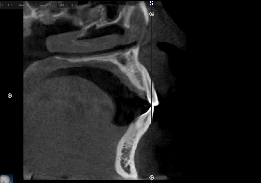
A case report by FKG UGM students published in the MKGK Journal of FKG UGM by Alhidayati Asymal, Menik Priaminiarti, Stefano Aditya Handoko, and Laura Susanti reported a patient with unilateral TMJ ankylosis accompanied by suspected fibrous dysplasia fibrous dysplasia of the mandibular ramus. CBCT was used as the primary diagnostic tool to evaluate the clinical presentation and bone anatomy. Sagittal and coronal CBCT images showed fusion of the left TMJ and anatomical TMJ structures that could not be identified. Histopathological examination (HPA) of the hypodense lesion revealed trabecular bone fragments suggestive of a fibro-osseous lesion. This emphasizes that CBCT is not only capable of demonstrating TMJ ankylosis, but also of revealing hypodense lesions in the mandibular ramus with characteristics of fibro-osseous lesions, which were subsequently confirmed by histopathology.
Radiological Analysis
- From sagittal and coronal CBCT images, a hypodense (radiolucent) lesion was observed in the mandibular ramus, and the left TMJ showed fusion (ankylosis) limiting mandibular mobility.
- TMJ anatomical structures could not be clearly identified on the affected side, indicating destruction or deformity due to the fibro-osseous lesion.
- CBCT provides three-dimensional data that allow evaluation of the extent of ramus involvement, severity, and potential spread to the joint or surrounding soft tissues.
Benefits of Early Diagnosis Through Radiology
- Earlier and More Accurate Intervention
With CBCT, fibro-osseous lesions can be identified before severe clinical manifestations occur, allowing for more conservative treatment planning or limited surgical resection. - Surgical and Oral Surgery Planning
Knowledge of lesion boundaries, involvement of surrounding tissues, and structural anatomy facilitates surgical planning (for example, bone resection, reconstruction, or TMJ correction). - Prognosis Prediction and Monitoring
Radiology assists in monitoring lesion growth, response to therapy, and potential complications such as progression toward the TMJ region or joint involvement. - Reducing Morbidity
Early diagnosis reduces the risk of facial deformity, functional disturbances (speech, mastication), and permanent joint damage.
***
CBCT plays a critical role as a radiological tool in the early identification of fibrous dysplasia of the jaw, particularly when it coexists with conditions such as TMJ ankylosis. The hypodense appearance of fibro-osseous lesions, clear bone destruction margins, and well-defined anatomical relationships enable more effective diagnosis and treatment planning. Early diagnosis through CBCT supports better care, reduces complications, and aligns with principles of health and sustainability.
References
MKGK, Alhidayati Asymal, Menik Priaminiarti, Stefano Aditya Handoko, Laura Susanti, CBCT examination in unilateral TMJ ankylosis with suspected fibrous dysplasia of the mandibular ramus, https://jurnal.ugm.ac.id/mkgk/article/view/82274
Author: Rizky B. Hendrawan | Photo: Freepik