Maxillofacial radiology is a critical field in the diagnosis and surgical planning of facial and jaw conditions. With advances in imaging technology (CT scan, CBCT, 3D imaging, navigation, and digital simulation), researchers and clinicians can precisely map the complex anatomy of the maxilla, mandible, facial bones, and surrounding structures.
In the context of complex maxillofacial fractures, research by Agung Hadi Wijanarko, a student of the Faculty of Dentistry, UGM, under the supervision of drg. Prihartiningsih, SU., Sp.BM(K), entitled “Integrated Management of Complex Maxillofacial Fractures with Malocclusion and Malunion”, emphasizes the importance of integrated diagnosis and planning for delayed fracture cases that often require correction of malocclusion and malunion. Radiological research plays a crucial role in supporting these steps.
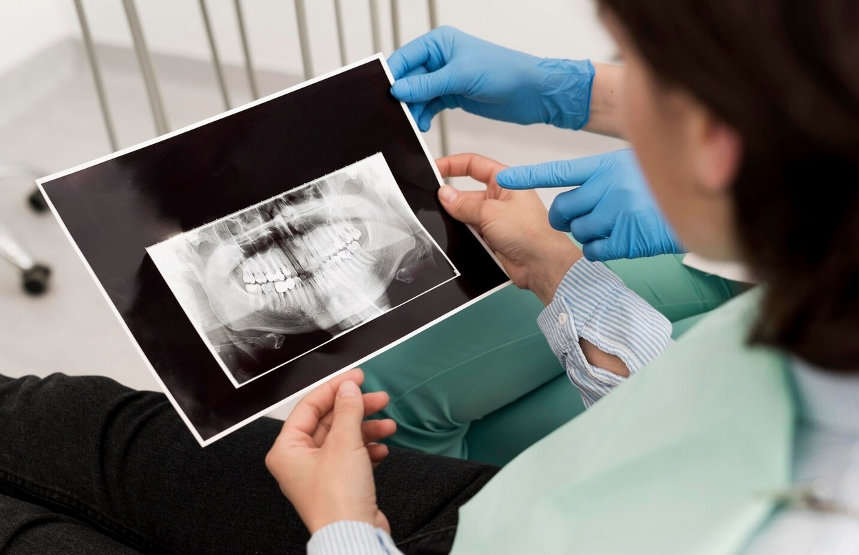
Role of Radiology in Maxillofacial Anatomy and Fractures
1. Diagnosis and fracture mapping
- Modalities such as CT and CBCT provide three-dimensional visualization of facial bone injuries, including displacement, bone fragments, sinus involvement, orbital floor involvement, and anatomical contours.
- Radiographic images allow surgeons to determine fracture types (e.g., Le Fort I, II, or combinations), malunion or malocclusion locations, and complex patterns not visible on conventional radiographs.
2. Integrated surgical planning
- Radiological data enable virtual simulation of bone reconstruction and jaw repositioning.
- Accurate anatomical mapping allows surgical teams to plan osteotomies, refracture procedures, repositioning, and fixation in a single-stage approach, as performed in UGM’s integrated management of complex maxillofacial fractures.
- Radiology helps determine optimal locations for plates, screws, and anchorage points.
3. Postoperative evaluation and healing monitoring
- Postoperative imaging (CT, CBCT) is used to evaluate bone position, jaw alignment, and osseous union.
- Radiological information helps assess residual malunion or the need for further correction.
Relation to UGM Research on Complex Maxillofacial Fractures
The UGM study described a delayed case of complex maxillofacial fractures (including Le Fort II and mandibular fractures) resulting in malocclusion and malunion. Integrated management was performed in a single surgical stage, including Le Fort I osteotomy, refracture, repositioning, and fixation. Six-month follow-up results showed:
- No infection or rejection of fixation devices.
- Progressive improvement in function and aesthetics.
The success of this integrated approach depended heavily on accurate facial bone anatomical data obtained through advanced radiological imaging.
Challenges and Future Directions in Maxillofacial Radiology
Beberapa tantangan dan peluang riset di bidang radiologi maksilofasial meliputi:
- High resolution and metal artifacts: implants and fixation plates can cause artifacts; research focuses on artifact reduction algorithms and advanced imaging techniques.
- AI integration and automated segmentation: development of AI algorithms to detect fractures, separate bone fragments, and predict optimal surgical plans.
- Image-guided intraoperative navigation: combining real-time imaging with surgical navigation for more precise fixation.
- Perfusion and vascular imaging: assessing blood supply to bone fragments to support better healing.
- Radiation dose reduction: optimizing protocols to minimize patient exposure while maintaining high diagnostic quality.
***
Radiological research focusing on maxillofacial anatomy is a critical foundation for diagnosis, surgical planning, and postoperative evaluation, especially in complex fracture cases. Advances in radiology (CT, CBCT, AI, navigation) continue to open new possibilities for more precise, safer, and personalized maxillofacial surgery.
References
WIJANARKO, Agung Hadi, drg. Prihartiningsih, SU.,Sp.BM(K), Integrated Management of Complex Maxillofacial Fractures with Malocclusion and Malunion, https://etd.repository.ugm.ac.id/penelitian/detail/36727
Author: Rizky B. Hendrawan | Photo: Freepik