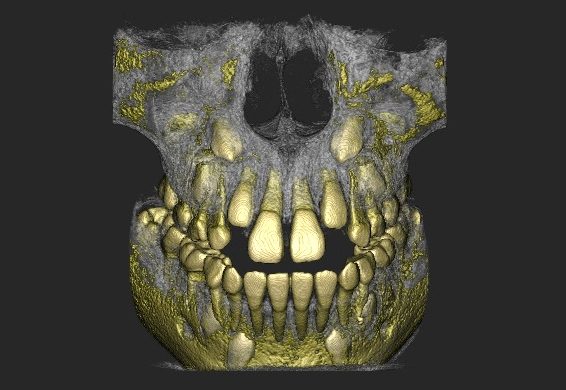
Odontoma is a benign odontogenic tumor composed of dental hard tissues and is a common cause of obstruction in the eruption pathway of permanent teeth, particularly the maxillary central incisors. A complex odontoma may block tooth growth and eruption, leading to impaction as well as aesthetic and functional problems, such as displacement of adjacent teeth and narrowing of the available space.
Research conducted by Lidya Noviana Arfiadi, Dr. drg. Cendrawasih Andusyana Farmasyanti, M.Kes., Sp.Ort(K), and Kuswayuning demonstrates that minimally invasive surgical techniques combined with orthodontic treatment can be an effective solution for managing complex odontomas that obstruct tooth eruption. The findings show that surgical removal of the odontoma followed by closed method exposure and light orthodontic traction can result in successful tooth eruption with satisfactory gingival contour and periodontal outcomes.
Case Description
- A 23-year-old female patient reported that the maxillary left central incisor (tooth 21) had not erupted since exfoliation of the primary tooth. Adjacent teeth (teeth 11 and 22) had shifted medially, causing narrowing of the edentulous space.
- Radiographic examination revealed radiopaque structures resembling small teeth along the eruption path of tooth 21, which were diagnosed as a complex odontoma.
Minimally Invasive Techniques Applied
The management involved a combination of surgical and orthodontic approaches:
- Orthodontic space preparation
Placement of a fixed orthodontic appliance to create sufficient mesiodistal space between teeth 11 and 22, allowing eruption of tooth 21. - Surgical removal of the odontoma
Surgical excision of the complex odontoma obstructing the eruption pathway. - Closed method exposure with light traction
After odontoma removal, tooth 21 was exposed using aclosed method exposureand guided into eruption with light orthodontic traction.
Outcomes and Advantages
- Six months after orthodontic appliance placement and exposure, tooth 21 successfully erupted into an ideal occlusal position.
- The labial gingival contour and periodontal tissues around the tooth root remained healthy.
- This procedure caused less damage to gingival and supporting tissues compared with more invasive exposure or resective surgical techniques.
Clinical Considerations
Several factors contributed to the success of the minimally invasive approach:
- A relatively favorable position and eruption direction of the impacted tooth.
- Complete root formation, allowing effective light orthodontic traction.
- Adequate inter-dental space created prior to exposure, preventing biological pressure on surrounding tissues and reducing the risk of periodontal damage.
***
Management of complex odontomas using a minimally invasive approach—consisting of odontoma surgery, closed method exposure, and light orthodontic traction following orthodontic space preparation—demonstrates a favorable prognosis. This approach enables ideal tooth eruption, preserves gingival and periodontal tissues, and minimizes surgical trauma.
References
Lidya Noviana Arfiadi, Dr. drg. Cendrawasih Andusyana Farmasyanti, M.Kes.Sp.Ort(K), Kuswayuning, Penatalaksanaan interdisipliner kasus impaksi gigi incisivus sentral maksila akibat obstruksi odontoma kompleks, https://jurnal.ugm.ac.id/mkgk/article/download/32000/19334
Author: Rizky B. Hendrawan | Photo: Freepik