
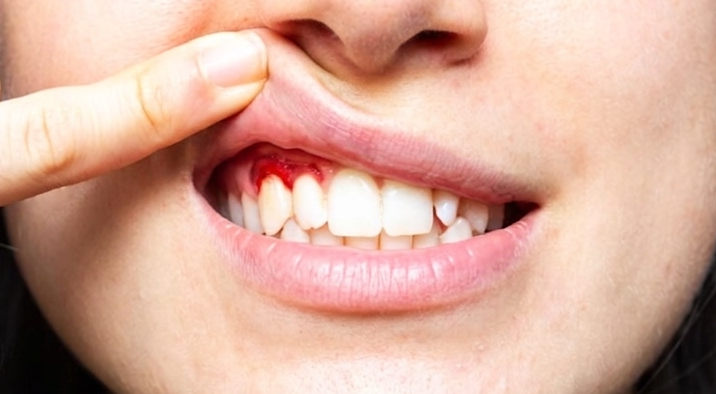
The thin membrane surrounding a fetus in the womb turns out to hold significant potential for healing gum wounds. This was the finding of research published in the International Journal of Oral and Maxillofacial Surgery in 2006 by drg. Margareta Rinastiti, M.Kes., Sp.KG(K)., Ph.D., from the Department of Conservative Dentistry, Faculty of Dentistry, Universitas Gadjah Mada. Conducted in Yogyakarta using a rabbit model, the study showed that transplantation of human amniotic membrane significantly accelerated gingival tissue recovery, with healing occurring much faster than natural healing without a wound covering.
The amniotic membrane is the innermost layer of the membranes that protect the fetus during pregnancy. It contains components of the basement membrane, growth factors, and proteinase-inhibiting compounds. This combination makes it unique: it does not merely provide protection, but also actively promotes tissue regeneration. Before this study, amniotic membranes had already been used as dressings for burns and skin ulcers. In ophthalmology, their transplantation had also been shown to accelerate corneal epithelialization and suppress inflammation.
Gingival tissue, or the gums, has unique characteristics. It naturally heals faster and produces less scarring than skin. However, under certain conditions, such as following periodontal surgery or soft-tissue reconstruction, additional support may still be needed.
The researchers divided 30 male rabbits aged 3–4 months into two groups. The first group was allowed to heal naturally after a biopsy wound was created on the buccal gingiva, the side of the gum facing the cheek, between the upper incisors and premolars. The second group received a covering consisting of five layers of human amniotic membrane, sutured with 5-0 Vicryl. The amniotic membrane was obtained from the Tissue Bank and Biomaterial Center of Dr. Soetomo Regional General Hospital, Universitas Airlangga, Surabaya, and had undergone a freeze-drying process. Gingival tissue samples were collected on days 1, 3, 5, 7, and 10 after the procedure and then processed for histological examination.
Four main parameters were evaluated: the number of PMN cells (polymorphonuclear inflammatory cells), the number of fibroblasts, the number of newly formed blood vessels, and epithelial thickness and collagen fiber density. The results were consistent and statistically significant. The group receiving the amniotic membrane showed fewer PMN cells from the first day, indicating a more controlled inflammatory response. New blood vessels had already formed by day 3, whereas they did not appear in the control group until day 5. Fibroblast numbers were higher from day 3 through day 10. The epithelial layer was significantly thicker from days 5 to 10. Collagen fibers were denser and better organized by day 10.
“Amniotic membrane transplantation appears to promote rapid epithelialization, granulation tissue formation, and collagen production, while suppressing inflammation, thereby accelerating gingival wound healing in rabbits compared with secondary healing,” the research team wrote in the publication.
This finding was not coincidental. The amniotic membrane contains growth factors such as bFGF, EGF, and TGF-β, which are known to stimulate fibroblast proliferation and new blood vessel formation. It also contains laminin, a substrate that facilitates epithelial cell attachment and migration to the wound area. In terms of inflammation, the amniotic membrane is thought to suppress PMN cell migration through several pathways: inducing inflammatory cell apoptosis, inhibiting the production of pro-inflammatory cytokines, and even preventing bacterial contamination because of its ability to adhere closely to the wound surface.
Interestingly, collagen fiber density in the amniotic membrane group was not merely greater but also better organized. In the control group, collagen fibers on day 10 still appeared immature and irregular. In contrast, in the amniotic membrane group, the fibers were already dense and neatly arranged, indicating more mature and functional tissue.
The clinical implications are promising. Cryopreserved human amniotic membrane has low immunogenicity because it expresses few MHC class II molecules and greater amounts of immunoregulatory molecules such as Fas ligand and HLA-G. This means that the risk of rejection by the recipient’s body, whether in the context of an allograft or xenograft, is relatively low, regardless of the patient’s genetic background. If these findings in rabbits can be confirmed in humans, amniotic membrane could potentially serve as an alternative biological material for periodontal surgery, gingival reconstruction, and the management of postoperative wounds in the oral cavity.
Author: drg. Achmad Zam Zam Aghasy, M.Kes., Annisa Dwi Noviyanti
Photo: FreePik
Source DOI: https://doi.org/10.1016/j.ijom.2005.09.012