
A finding from the Faculty of Dentistry, Universitas Gadjah Mada, has shown that a hydrogel-based scaffold material combined with carbonate apatite can significantly promote fibroblast cell growth. The study, published in the Journal of Dentistry Indonesia, was conducted by drg. Ivan Arie Wahyudi, M.Kes., Ph.D., from the Department of Biomedical Sciences, Faculty of Dentistry, Universitas Gadjah Mada, together with Lea M. Nurwadji, with the aim of finding a more effective solution for bone reconstruction in patients with bone damage caused by surgical procedures or degenerative diseases. The results showed that on the third day of observation, fibroblast proliferation increased significantly compared with the control group.
Damaged Bone and Unresolved Challenges
Bone is more than simply the body's structural framework. It supports posture, protects vital organs, serves as a site for blood cell production, and stores calcium and phosphate reserves. When bone sustains extensive damage—for example, following tumor removal surgery, trauma, or certain diseases—the body is not always able to repair it on its own.
In dentistry, loss of alveolar bone, the bone that supports the teeth, can lead to tooth loss, impaired chewing, and difficulties with denture placement. Common treatments include bone grafting and the placement of barrier membranes through Guided Tissue Regeneration (GTR) or Guided Bone Regeneration (GBR) techniques.
However, each approach has its limitations. Autografts, which use bone harvested from the patient's own body, are considered the gold standard, but their availability is limited and they create an additional wound at the donor site. Bone obtained from other human donors or animals carries a risk of disease transmission. Synthetic materials, meanwhile, may trigger adverse tissue reactions. GTR techniques also have limited indications because they are prone to failure when wound closure is incomplete.
This is where tissue engineering offers a new path forward.
A Miniature Scaffold That Mimics the Cellular Environment
Tissue engineering operates on a simple principle: provide a “home” where cells can grow, develop, and form new tissue. That home is known as a scaffold.
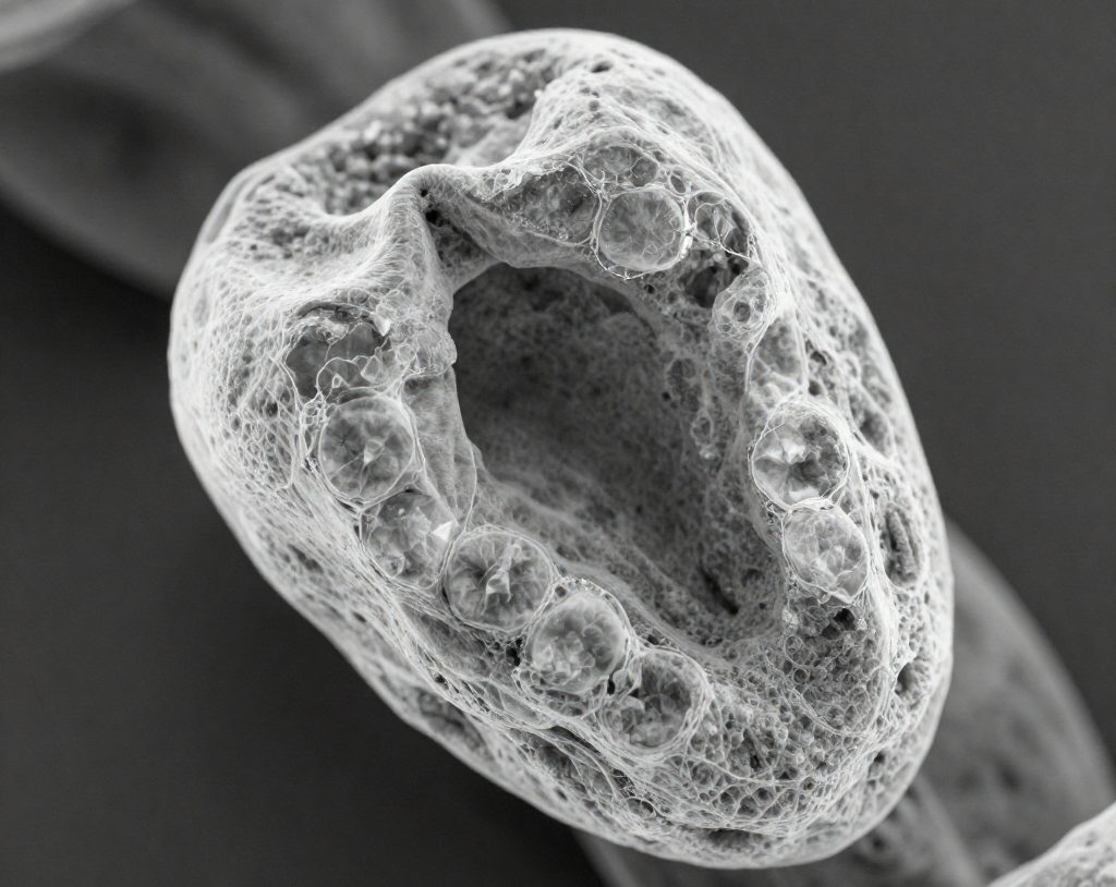
Hydrogel is one promising scaffold material. Its structure resembles the extracellular matrix, the natural environment in which the body's cells live and communicate. Hydrogels are hydrophilic, capable of absorbing water, and generally well tolerated by biological tissues. When combined with carbonate apatite (CHA), a mineral whose chemical composition resembles that of natural bone mineral, the resulting scaffold is expected to be not only mechanically suitable but also biologically compatible.
What distinguishes this study from previous research is the method used to produce the hydrogel: non-freeze-dried, meaning that it does not undergo a freeze-drying process. Conventional freeze-drying produces pores in the scaffold, but their size can be difficult to control. The non-freeze-dried method produces smaller pores, which may actually be advantageous because they are more effective at preventing connective tissue from entering and interfering with new bone formation.
Counting Cells One by One
To test the effectiveness of this material, the researchers used human fibroblasts obtained from circumcision tissue. Fibroblasts were selected because they are easy to culture in the laboratory, grow rapidly, are non-tumorigenic, and have the potential to differentiate into osteoblasts, the cells responsible for bone formation.
The cells were divided into three groups: fibroblasts alone without treatment, fibroblasts with non-freeze-dried hydrogel, and fibroblasts with non-freeze-dried hydrogel-CHA. Each group was seeded at a density of 2 × 10⁴ cells per milliliter in 96-well culture plates. Cell growth was observed under a microscope and quantified using the MTT assay, a standard method for measuring the number of viable cells, on days 1, 2, and 3.
The results were quite clear. On the first day, there were no significant differences among the groups. However, by the third day, the group treated with hydrogel-CHA showed a substantially higher number of fibroblasts than the untreated group, with a statistically significant difference (p < 0.05).
“Inorganic and organic composition of the hydrogel-CHA can stimulate fibroblast proliferation more rapidly.”
The explanation makes biological sense. The calcium-phosphate components of CHA are thought to increase the swelling capacity of the hydrogel, thereby expanding the surface area available for cells to attach and proliferate. The porous scaffold also allows oxygen, nutrients, and metabolic products to diffuse in and out, creating a microenvironment that supports cell growth.
From the Laboratory to the Clinic
Of course, the journey from laboratory findings to clinical application is still a long one. This study was conducted in vitro, meaning in a culture dish rather than inside the human body. The observation period was also limited to three days, in accordance with previous research showing that fibroblast morphology does not change significantly within that period.
Nevertheless, these findings establish an important foundation. Non-freeze-dried hydrogel-CHA was shown to be non-toxic to cells and even actively promoted their growth. This is a basic requirement that must be met before a material can be considered for further testing in animals and eventually humans.
For patients who lose the bone supporting their teeth because of severe periodontal disease or surgical procedures, materials like this offer hope. One day, dentists may be able to repair bone defects not by harvesting bone from another part of the body, but by using an intelligent synthetic scaffold that invites the body's own cells to rebuild what has been lost.
Authors: Achmad Zam Zam Aghasy, DDS, M.Kes.; Hazra Alifia Muharam
Photo: Freepik
Sumber DOI: https://doi.org/10.14693/jdi.v21i3.226