
The bacterium Enterococcus faecalis has long been a major challenge in root canal treatment. This bacterium is difficult to eliminate: it can penetrate deep into dentinal tubules, form a protective layer known as a biofilm, and survive exposure to conventional antiseptic agents, including calcium hydroxide, the most commonly used root canal medicament worldwide. Now, a laboratory study from the Department of Conservative Dentistry, Faculty of Dentistry, Universitas Gadjah Mada (UGM), has found that chitosan nanoparticles—materials derived from shrimp and crab shells—can outperform chlorhexidine and glycerin as vehicles for calcium hydroxide in killing the bacterium, while also releasing calcium ions and producing a higher pH. The study was published in the Journal of Conservative Dentistry in September–October 2022 and involved Prof. drg. Diatri Nari Ratih, M.Kes., Ph.D., Sp.KG(K), Dr. drg. Ema Mulyawati, M.S., Sp.KG(K), and drg. Henytaria Fajrianti, Sp.KG.
Root canal treatment, commonly referred to by the public as “nerve treatment,” is not simply a procedure for cleaning the infected contents of a tooth. It is a highly precise sterilization process. Dentists must ensure that bacteria within the root canal are thoroughly eliminated before the canal is permanently sealed.
The problem is that root canal anatomy is not as simple as a straight tube. There are branches and narrow spaces that are difficult for mechanical instruments to reach. This is where bacteria can hide and multiply. Although mechanical cleaning can significantly reduce bacterial populations, residual colonies can lead to treatment failure, recurrent infection, abscess formation, or even tooth loss.
To address this problem, dentists use intracanal medicaments—medications placed inside the root canal between treatment visits and allowed to act for one to two weeks. Calcium hydroxide has been the primary choice for decades. It works by dissociating into calcium and hydroxyl ions, creating a highly alkaline environment that is lethal to bacteria. However, it has one crucial weakness: calcium hydroxide alone is not sufficiently effective against E. faecalis. This bacterium can survive even under highly alkaline conditions.
In clinical practice, calcium hydroxide is not used as a pure, dry substance. It must be mixed with a solvent, scientifically referred to as a vehicle, to form a paste that can be applied inside the root canal using a specialized instrument called a lentulo spiral. The vehicle is not simply a diluent. It determines how quickly calcium hydroxide dissociates, how many ions are released, and how effectively the medication kills bacteria. Two commonly used vehicles are glycerin and chlorhexidine gluconate (CHX).
Glycerin is easy to use because its viscous consistency facilitates application into the root canal. However, this viscosity is also its weakness. Calcium and hydroxyl ions are released very slowly, resulting in relatively weak antibacterial activity, particularly against E. faecalis. CHX is superior in terms of bactericidal activity. Its cationic molecules bind to negatively charged bacterial cell walls and damage them from within. However, CHX also presents a problem as a vehicle: calcium hydroxide-CHX paste can be difficult to distribute evenly throughout the entire root canal, making its effectiveness inconsistent.
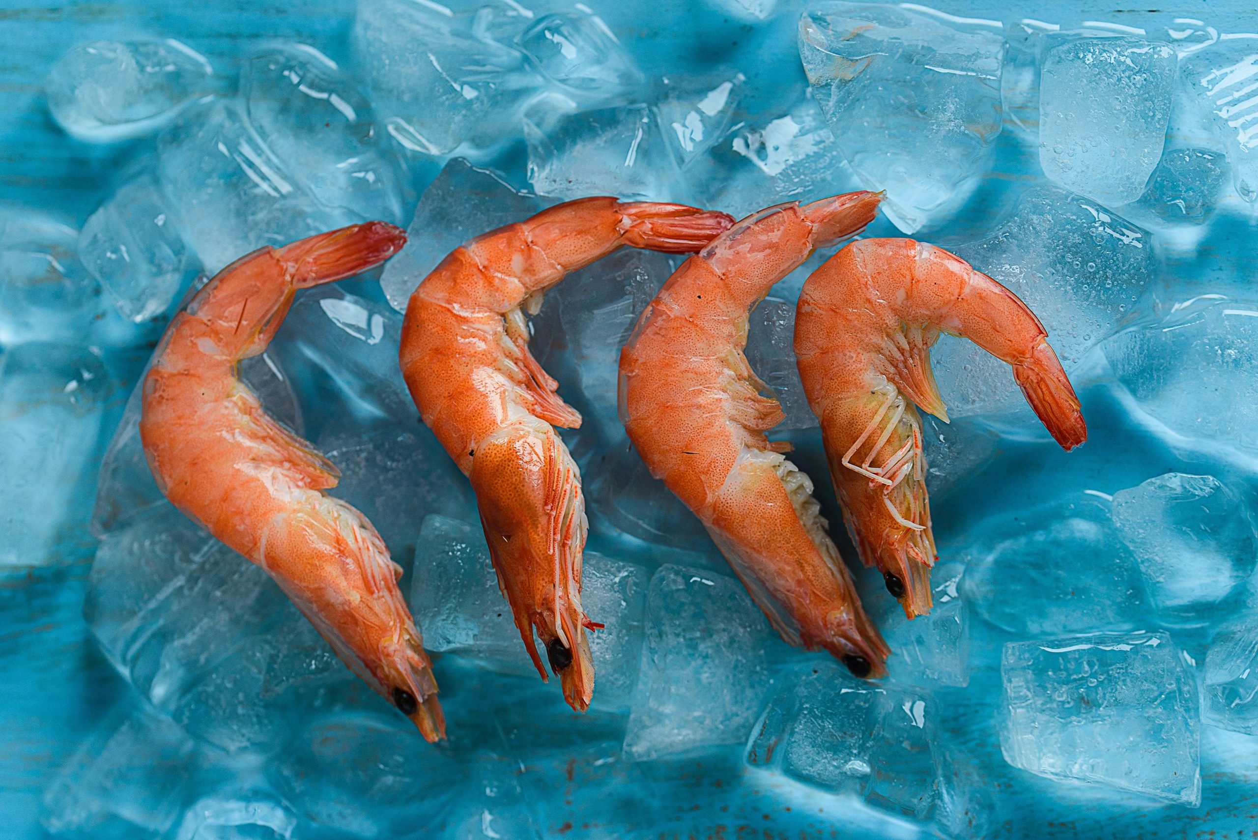
Chitosan is not a new material. It has long been studied in biomedical research as a natural polymer obtained through the deacetylation of chitin, the main component of crustacean shells such as shrimp, crab, and lobster shells. In dentistry, chitosan has been investigated for various applications, ranging from root canal irrigation agents to dentin remineralization materials. What makes chitosan nanoparticles particularly interesting is their extremely small size. Because they are smaller, their surface area per unit of mass is much greater than that of conventional chitosan. This provides more points of contact with bacterial cell surfaces and, consequently, greater bactericidal activity.
Its mechanism of action is also unique. Chitosan is positively charged, whereas bacterial cell surfaces are negatively charged. The two attract each other like magnets. Once chitosan attaches to the bacterial cell membrane, it alters membrane permeability, causing essential intracellular components to leak out and ultimately killing the bacteria. A second mechanism involves chitosan inducing the production of reactive oxygen species, which damage bacterial proteins and DNA. In addition, chitosan is aqueous, meaning that it facilitates faster and more efficient hydroxyl-ion release than glycerin, which is highly viscous.
The experimental study was designed to systematically compare the three vehicles. Three parameters were evaluated: antibacterial effectiveness against E. faecalis, calcium-ion release, and changes in pH. All parameters were measured at two time points: 7 and 14 days.
For the antibacterial test, the agar diffusion method was used. E. faecalis ATCC 29212 was cultured on agar media, after which 6-mm-diameter wells were prepared and filled with the respective calcium hydroxide pastes. Following 24 hours of incubation, the inhibition zones—the clear areas surrounding the wells indicating bacterial death—were measured using a vernier caliper.
For calcium-ion release and pH measurements, 24 single-rooted premolar teeth with prepared root canals were used. Each tooth was immersed to one-third of its apical portion in distilled water and then stored in an incubator. The immersion solution was collected periodically and analyzed using atomic absorption spectrometry for calcium ions and a pH meter for pH measurements.
The results of the three tests showed that chitosan nanoparticles performed best across all parameters. The bacterial inhibition zone in the chitosan group was significantly larger than that in the glycerin group (P < 0.05), although the difference compared with CHX was not statistically significant. For calcium-ion release and pH, chitosan outperformed both glycerin and CHX at both time points (P < 0.05). More interestingly, the pH in the chitosan group continued to increase from day 7 to day 14, indicating that chitosan was capable of sustaining hydroxyl-ion release over a longer period.
These findings have considerable practical significance. Dentists who wish to maximize antibacterial activity have traditionally mixed calcium hydroxide with CHX, a combination that is indeed effective but has the disadvantage of uneven distribution. Chitosan nanoparticles offer a promising alternative: better distribution than CHX due to their more manageable consistency, antibacterial activity that is comparable to or greater than that of CHX, and greater calcium-ion release.
Calcium ions themselves are not merely indicators of medication activity. They play an active role in the healing process by promoting extracellular matrix mineralization, supporting tissue repair in the periapical region, and even activating adenosine triphosphate (ATP), which plays an important role in hard-tissue mineralization.
Chitosan also has chelating properties toward calcium ions. It can bind calcium ions to its surface, allowing their release to occur in a more controlled and stable manner. This makes it an interesting candidate for cases requiring long-term intracanal medication.
Of course, this study was conducted at the laboratory level. Clinical trials involving actual patients are still necessary before chitosan nanoparticles can be recommended as a new clinical standard. Nevertheless, as preliminary evidence, the findings are sufficiently compelling to encourage further research into formulation, biological safety, and effectiveness under more complex root canal conditions.
Author: drg. Achmad Zam Zam Aghasy, M.Kes., Annisa Dwi Noviyanti
Photo: FreePik
Source DOI: https://doi.org/10.4103/jcd.jcd_242_22