
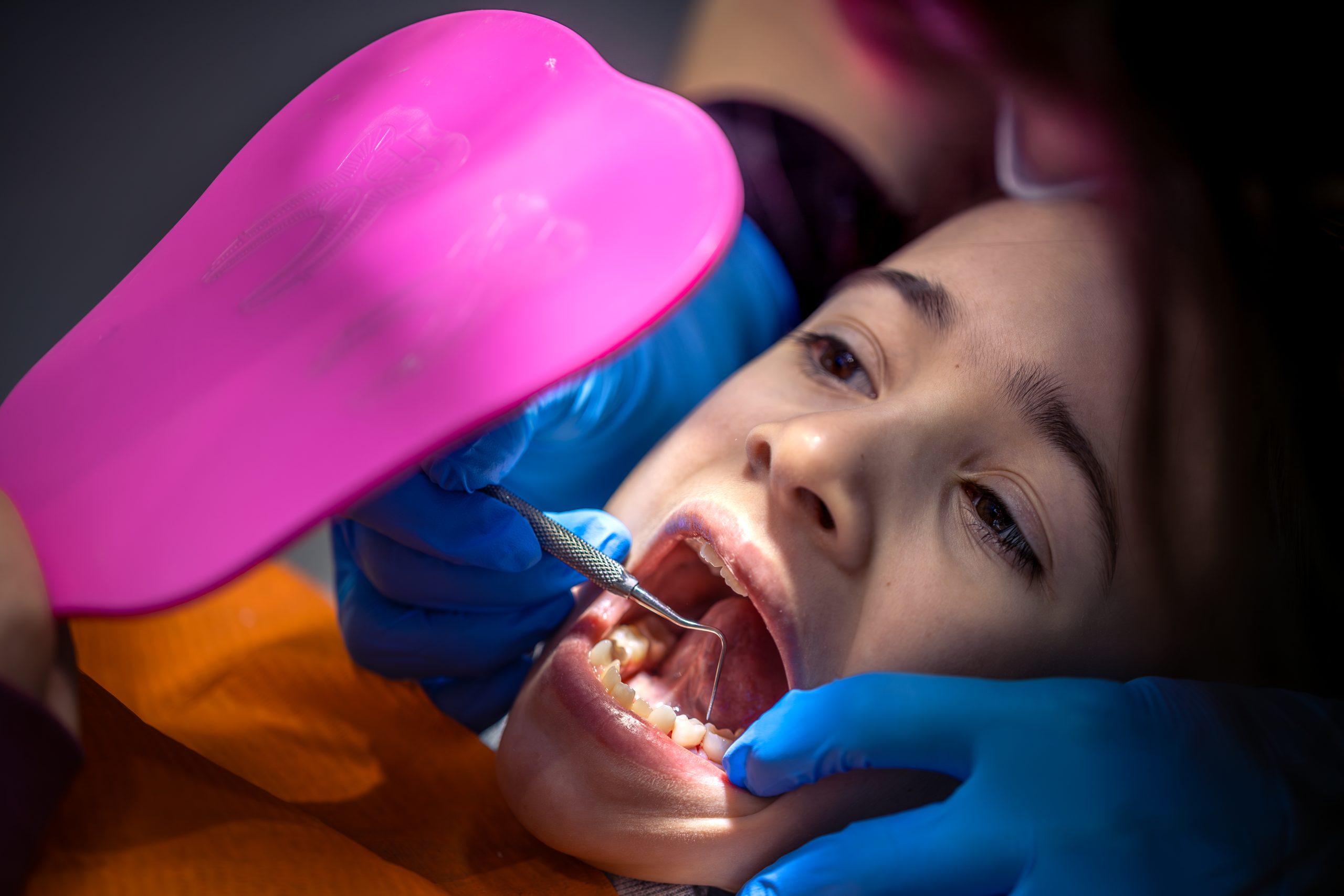
The 2.5-year-old boy arrived at the emergency department of Temanggung Regional Hospital with his face covered in blood. Thirty minutes earlier, the motorcycle he was riding with his grandfather—sitting in front of the driver without a helmet—had collided with a stationary trailer truck. His lower jaw was fractured. His primary teeth had been knocked out. And deep inside his still-developing jawbone lay the tiny tooth buds of permanent teeth that had never yet seen the light of day.
This was the challenge faced by the oral and maxillofacial surgery team from the Faculty of Dentistry, Universitas Gadjah Mada: saving a toddler’s jaw without sacrificing the future of his permanent teeth.
A Fracture That Was More Than a Broken Bone
Clinical examination revealed a serious condition. The child’s face appeared asymmetrical. Inside the mouth, there was a laceration between teeth 71 and 81, one avulsed primary tooth, discontinuity of the mandible with the right segment displaced lingually and superiorly, and a prominent sublingual hematoma.
A three-dimensional CT scan confirmed the diagnosis: a mandibular symphysis fracture with significant displacement of the fragments. In adults, managing an injury like this is relatively straightforward. But in a 30-month-old child, doctors are dealing with completely different anatomy. A child’s cortical bone is thinner and more fragile. Primary teeth have short crowns with maximum convexity in the gingival third, making fixation wires prone to slipping. Most importantly, along the fracture line were the developing buds of the permanent incisors and canines, which were already at the apposition stage—the critical phase of crown and root formation.
According to Alqahtani et al. (2010), by 2.5 years of age, the mandibular permanent incisor and canine tooth buds have reached the apposition stage. Iatrogenic trauma during this phase can interfere with permanent tooth eruption and may even affect the alveolar process and jaw arch development permanently.
A Splint Born on the Operating Table
The team, consisting of resident drg. Lina Mariana, drg. Bramasto Purbo Sejati, Sp.BMM, and Prof. drg. Poerwati Soetji Rahajoe, Sp.BMM(K), decided against the conventional approach. Open reduction with a titanium plate was rejected because of the risk of damaging the developing tooth buds. Arch-bar fixation was not feasible because the primary teeth had crowns that were too short. Intermaxillary fixation (IMF) was also avoided because restricting food intake in pediatric patients could lead to nutritional deficiencies and significant weight loss.
The team instead chose a thermoformed splint—a device more commonly used as a mouthguard for patients with bruxism or as a bleaching tray—but this time fabricated directly on the operating table while the patient was under general anesthesia.
The process began by taking an impression of the jaw using alginate and dental stone. The fractured segments were separated and manually reduced on the plaster model to simulate the position that would be achieved clinically.
A transparent 1-mm-thick thermoplastic sheet was then pressed over the model using a vacuum-forming machine. The edges of the splint were trimmed to approximately 2 mm below the free gingival margin to provide additional mechanical retention. After the fracture segments were reduced bidigitally in the operating room, the splint was placed and secured using four circum-mandibular wires made of 0.5-mm stainless steel wire—two on each side of the fracture line.
“Treatment of pediatric mandibular fractures using a thermoformed splint fixed with circum-mandibular wiring provides good results and is an effective, minimally invasive approach for preventing iatrogenic injury to anatomical structures surrounding the fracture line.” — Lina Mariana, Bramasto Purbo Sejati, Poerwati Soetji Rahajoe, in the proceedings of the 7th Dental Seminar, Universitas Muhammadiyah Surakarta
Two Months, One Year, and a Healed Jaw
Seven days after surgery, extraoral edema was still visible, but the child appeared calm. There was no pain and no sign of infection. He could open and close his mouth normally—an important detail for preventing temporomandibular joint ankylosis, a serious complication that can permanently restrict jaw movement.
At follow-up visits two weeks, three weeks, one month, and two months after surgery, the condition continued to improve. The splint remained securely fixed, and the occlusion was normal. After two months, the splint and wires were removed under general anesthesia. A postoperative three-dimensional CT scan showed encouraging results: the fracture fragments had been perfectly reduced, the alignment of the lower border of the mandible had been restored, and—most importantly—evidence of osseous healing was visible in the mandibular symphysis region.
Evaluations at six months and one year after surgery showed normal extraoral conditions. There was no pain, infection, or limitation of jaw movement. Occlusion remained normal.
The main advantage of this method, as discussed in the case report, lies in the splint’s uniform thickness. There was no premature contact that could interfere with the bite. The transparency of the material allowed clinicians to monitor tooth adaptation externally. Its three-dimensional flexibility also permitted physiological tooth mobility without transferring unwanted orthodontic forces to developing teeth.
When an Accident Could Have Been Prevented
But this case report is not only about surgical technique. Near the end of the paper, almost like a footnote, appears a statement that is actually central to the entire story: public education is needed to promote optimal head protection for road users, including children, and to ensure that children are positioned in safer areas when riding motorcycles.
The 30-month-old boy was sitting in front of the driver, without a helmet, on the same road as large trucks. A significantly displaced mandibular symphysis fracture was the consequence of a common—and preventable—form of negligence in Indonesia.
For now, his permanent tooth buds were spared. But how many other children are not so fortunate, and how many are not treated by teams sufficiently trained to select the right approach?
Authors: Nanda Ayu, drg. Achmad Zam Zam Aghasy, M.Kes.
Photo: Freepik