
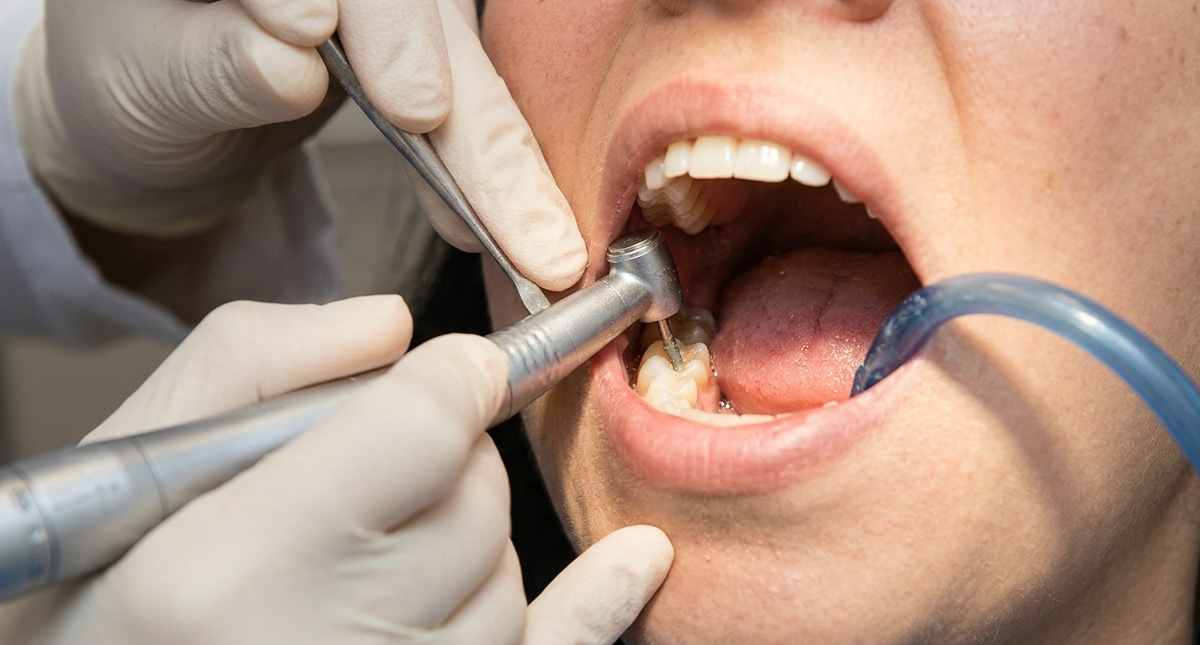
The sharp sensation that appears after a tooth filling is not merely a common side effect that can be ignored. A study published in the journal Dental Materials (2007) revealed the mechanisms behind this complaint, which is frequently experienced by patients. Prof. drg. Diatri Nari Ratih, M.Kes., Ph.D., Sp.KG(K), together with colleagues from the Department of Restorative Dentistry, School of Dental Science, University of Melbourne, Australia, investigated what actually occurs inside the tooth during composite resin restoration, a plastic-based filling material that is currently one of the most widely used restorative materials. They discovered that every stage of the filling procedure triggers fluid movement within the dentin and physical displacement of the tooth cusps. These movements continue even after the restoration has been completed.
Dentin is the hard layer beneath the enamel that surrounds the tooth. Within dentin are millions of microscopic channels called dentinal tubules, which contain fluid. This fluid plays a crucial role in tooth sensitivity: whenever the fluid moves, nerve endings inside the tooth become stimulated. This phenomenon is known as the hydrodynamic theory and serves as the foundation of the research conducted by the team.
In the study, ten extracted human maxillary premolars were prepared with MOD cavities (mesio-occluso-distal cavities), which extend across the mesial, occlusal, and distal surfaces of the tooth and represent one of the most extensive cavity preparations commonly encountered in clinical practice. Each tooth was connected to a fluid-flow measuring device called Flodec, while special sensors (DCDT) were attached to both sides of the tooth cusps to detect physical displacement as small as one micrometer. Both instruments operated simultaneously, recording what occurred inside the tooth during each stage of the filling procedure.
Composite resin restoration involves several sequential steps: etching with phosphoric acid to clean the dentin surface, rinsing and drying, application of bonding material (adhesive), and light curing to harden the restorative material.
During air drying, rapid and substantial contraction of the tooth cusps occurred, with an average displacement of 4.3 micrometers. Fluid within the dentinal tubules moved outward from the tooth at a much higher rate than under normal conditions. This occurred because the air stream caused evaporation of fluid on the surface of the tubules, pulling fluid from the inside toward the outside.
The stage that produced the greatest response was light curing, which hardens the composite resin. As the resin polymerizes, the material undergoes shrinkage. This shrinkage creates mechanical stress that pulls the tooth cusps inward. Cusp contraction during light curing reached an average of 8 micrometers, while dentinal fluid moved inward toward the pulp or the tooth’s nerve chamber at the highest rate recorded throughout the procedure, approximately 22.9 nL/second/cm².
“The large and rapid fluid movement during restoration, as well as the prolonged outward fluid flow after light curing, has implications for postoperative sensitivity.” — Ratih, Palamara, and Messer, Dental Materials (2007)
This finding is the most clinically relevant aspect of the study. Approximately one to two minutes after light curing was completed, dentinal fluid flow reversed direction, moving slowly but continuously outward, and continued for at least 15 minutes (the entire duration of the observation period). The fluid flowed outward through microscopic gaps between the filling material and the dentin surface.
Observations using Scanning Electron Microscopy (SEM) confirmed the presence of these gaps. In epoxy replicas, the spaces between dentin and composite resin measured between 3 and 20 micrometers. These gaps formed because polymerization shrinkage of the resin caused separation between the restorative material and the cavity walls.
These gaps create an open pathway between the oral cavity and the pulp–dentin complex. According to the researchers, continuous outward fluid movement through these spaces is likely one of the major causes of pulpal damage and the sensitivity experienced by patients after restoration.
The study also revealed that the relationship between mechanical stress and dentinal fluid movement is more complex than previously assumed. Only during light curing did the direction of fluid movement and cusp contraction occur in the same direction, with both moving inward. During other stages, such as drying and after light curing, fluid movement and cusp displacement occurred in opposite directions. This indicates that mechanical stress is only one of many factors influencing dentinal fluid movement. Thermal effects, evaporation, and osmotic factors also contribute simultaneously.
This research provides a strong scientific foundation for understanding why postoperative sensitivity occurs after dental fillings, while also opening opportunities for the development of improved restorative materials and techniques. Materials with lower polymerization shrinkage, gentler drying techniques, and stronger adhesive systems may theoretically reduce excessive fluid movement during and after restoration procedures.
For patients, this understanding confirms that sensitivity after a filling does not necessarily indicate treatment failure. Instead, it represents a predictable biological response that can now be explained scientifically down to the micrometer level.
Author: drg. Achmad Zam Zam Aghasy, M.Kes., Annisa Dwi Noviyanti
Photo: FreePik
Source DOI: https://doi.org/10.1016/j.dental.2006.11.029