
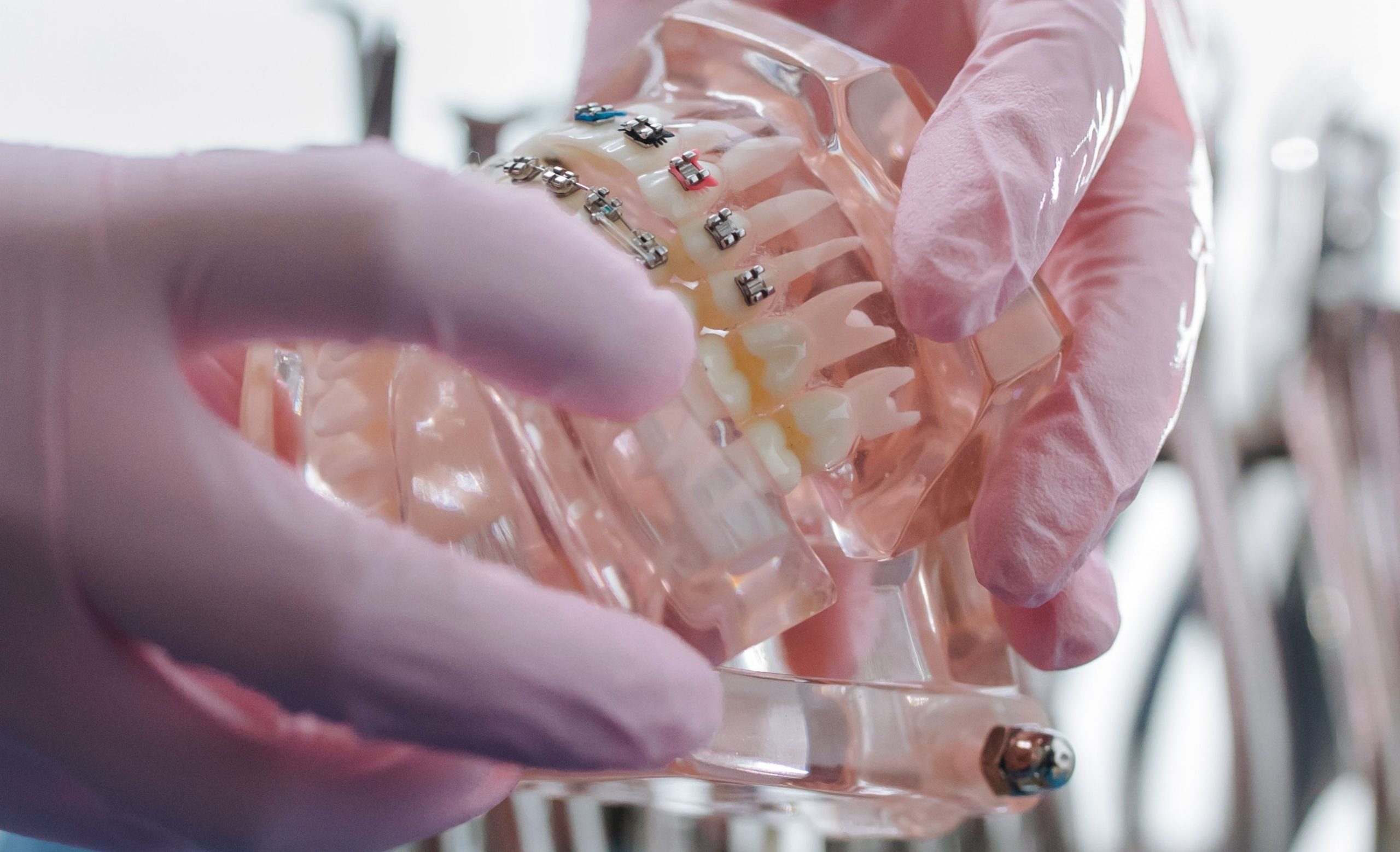
Imagine a patient sitting in an orthodontic chair, with delicate wires attached to their teeth and small elastic bands gently guiding the upper and lower jaws into a more harmonious position. Month after month passes. When the braces are finally removed, what changes is not only the alignment of the teeth—the shape of the face also undergoes a transformation that is subtle, measurable, and statistically significant.
This is the main finding of a study published in the Insisiva Dental Journal (Vol. 7 No. 1, May 2018), authored by M. Shulchan Ardiansyah together with two researchers from the Department of Orthodontics, Faculty of Dentistry, Universitas Gadjah Mada (UGM): Pinandi Sri Pudyani and Dr. Sri Suparwitri, DDS, S.U., Orthodontic Specialist. The study examined an aspect that is often overlooked by patients: how fixed orthodontic treatment using the Begg technique changes facial vertical proportions in measurable ways.
When Deep Bite Becomes a Facial Problem
Class II Division 1 malocclusion—a condition in which the upper teeth protrude excessively forward with increased overjet—is not merely a dental aesthetic issue. This condition is often accompanied by a deep bite, where the crowns of the upper incisors overlap half or more of the crowns of the lower incisors. As a result, anterior facial height becomes reduced, facial proportions appear unbalanced, and the condition may affect not only self-confidence but also a person’s psychological well-being.
The study analyzed 17 pairs of lateral cephalograms—profile X-ray images of the head taken before and after treatment. The participants, aged 18–35 years, were divided into two groups: Group I: Patients with an overjet of 2–4 mm and Group II: Patients with an overjet of 4.1–6 mm. Using these cephalograms, the researchers performed tracing and measurements of two main variables: the occlusal plane angle and the Facial Height Index (FHI), which represents the ratio between posterior and anterior facial height.
Small Numbers, Significant Changes
The measurements showed changes in the occlusal plane angle ranging from 2° to 5° after treatment, while facial height index changes ranged from 0.018 to 0.084 mm. These numbers may appear small, but in the context of human facial geometry, even minor changes can significantly alter facial appearance and profile.
More importantly, the direction of the relationship was notable. Spearman correlation analysis showed a significant negative correlation between changes in occlusal plane angle and changes in facial height index in both Group I (correlation coefficient -0.810) and Group II (correlation coefficient -0.696). This means that the greater the change in the occlusal plane angle during treatment, the smaller the change in facial height index—and vice versa.
“Control of facial vertical dimensions through mechanical appliances is very important to ensure that the expected treatment objectives can be achieved.”
This statement is not merely theoretical. It represents a fundamental principle that determines the success of the entire orthodontic treatment process.
The Begg Technique and the Secret Behind Arch Wires
The Begg technique, first introduced in 1956, operates through a differential force mechanism. The anchorage bend in the archwire, combined with vertical forces generated by Class II intermaxillary elastics, causes molars to undergo extrusion while incisors experience simultaneous retraction and intrusion. This combined effect opens the bite and produces changes in facial vertical dimensions.
In Group II, which had greater overjet, the average facial height index was lower compared with Group I. This finding is consistent with the mechanism of the Begg technique: the greater the correction required, the greater the tooth movement, and the greater the potential impact on anterior facial height. The Mann–Whitney test confirmed significant differences between the two groups (P < 0.05) for both measured variables.
Facial Proportion as a Determining Factor in Treatment Success
This study highlights that the facial height index is not merely an additional number in cephalometric analysis. An FHI value ranging from 0.55 to 0.85—with an ideal average of 0.70—serves as an indicator of mandibular rotation during treatment. When FHI increases, the mandible rotates upward and forward. When it decreases, the mandible moves downward and backward. According to the study, cases with values outside this range should be considered for combined orthodontic-surgical treatment.
For orthodontic practitioners, these findings deliver an important message: placing braces is one thing, but understanding how each mechanical force alters facial proportions in three dimensions is something entirely different. Patients sitting in the orthodontic chair are unknowingly undergoing a precise reconstruction of facial geometry—guided by wires as thin as a strand of hair and elastics as small as rubber bands.
Authors: Nanda Ayu; drg. Achmad Zam Zam Aghasy, M.Kes.
Photo: Pexels