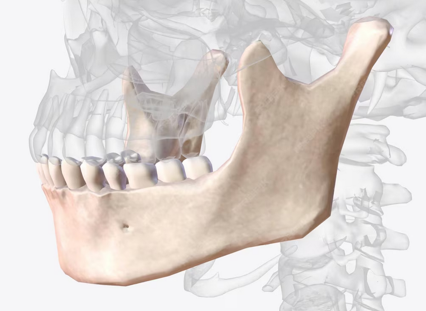
Temporomandibular Joint Disorders (TMD) refer to conditions affecting the temporomandibular joint (TMJ) and its supporting structures, which may cause pain, limited mandibular movement, and altered stomatognathic function. Accurate diagnosis of TMD requires a multidisciplinary approach, one of which involves imaging techniques that provide detailed visualization of both hard and soft tissues of the TMJ.
The Role of Imaging in TMD Diagnosis
Clinical examination alone is insufficient for comprehensive TMJ evaluation. Many pathological conditions—such as disc displacement, joint degeneration, and inflammation—cannot be directly observed without imaging. Therefore, imaging plays a crucial role in:
- Confirming clinical diagnose.
- Differentiating types of TMJ dysfunction
- Treatment planning
- Evaluating therapeutic outcomes
Various imaging modalities may be used, including conventional radiography, computed tomography (CT), cone-beam CT, and magnetic resonance imaging (MRI). Each modality has its advantages; however, MRI is considered the gold standard for evaluating soft tissue structures, particularly the articular disc.
Magnetic Resonance Imaging (MRI) as the Gold Standard
MRI provides high-resolution visualization of the articular disc and surrounding soft tissues without exposure to ionizing radiation. It is particularly effective in identifying:
- Disc position in closed- and open-mouth conditions.
- Degenerative joint changes
- Joint effusion and signs of inflammation
- Incongruence between the condyle and the fossa
These insights enable clinicians to establish more accurate diagnoses and select the most appropriate treatment strategies.
Related Research
Research conducted by an FKG UGM student, Wawan Suridwan, under the supervision of Prof. Dr. drg. Haryo Mustiko D., MS., Sp.Pros(K), entitled “The Use of Mouthguards in Early Treatment of Temporomandibular Disorders Using a Magnetic Resonance Imaging Approach,” demonstrated that MRI can effectively evaluate the outcomes of mouthguard therapy by visualizing changes in disc position and improvements in joint function during treatment.
***
Imaging is a critical component in the diagnosis and management of TMD. Selecting appropriate imaging techniques—with MRI as the primary modality for soft tissue assessment—enables accurate detection, effective treatment planning, and comprehensive evaluation of therapeutic outcomes. As an integral part of modern dental practice, imaging for TMD continues to evolve toward more precise and patient-safe approaches.
References
SURIDWAN, Wawan, Prof Dr. drg. Haryo Mustiko D., MS., Sp Pros(K), Penggunaan mouthguard pada perawatan awal kasus temporomandibular dissorders dengan pendekatan pemeriksaan magnetic resonance imaging, https://etd.repository.ugm.ac.id/penelitian/detail/37915
Author: Rizky B. Hendrawan | Photo: Freepik