
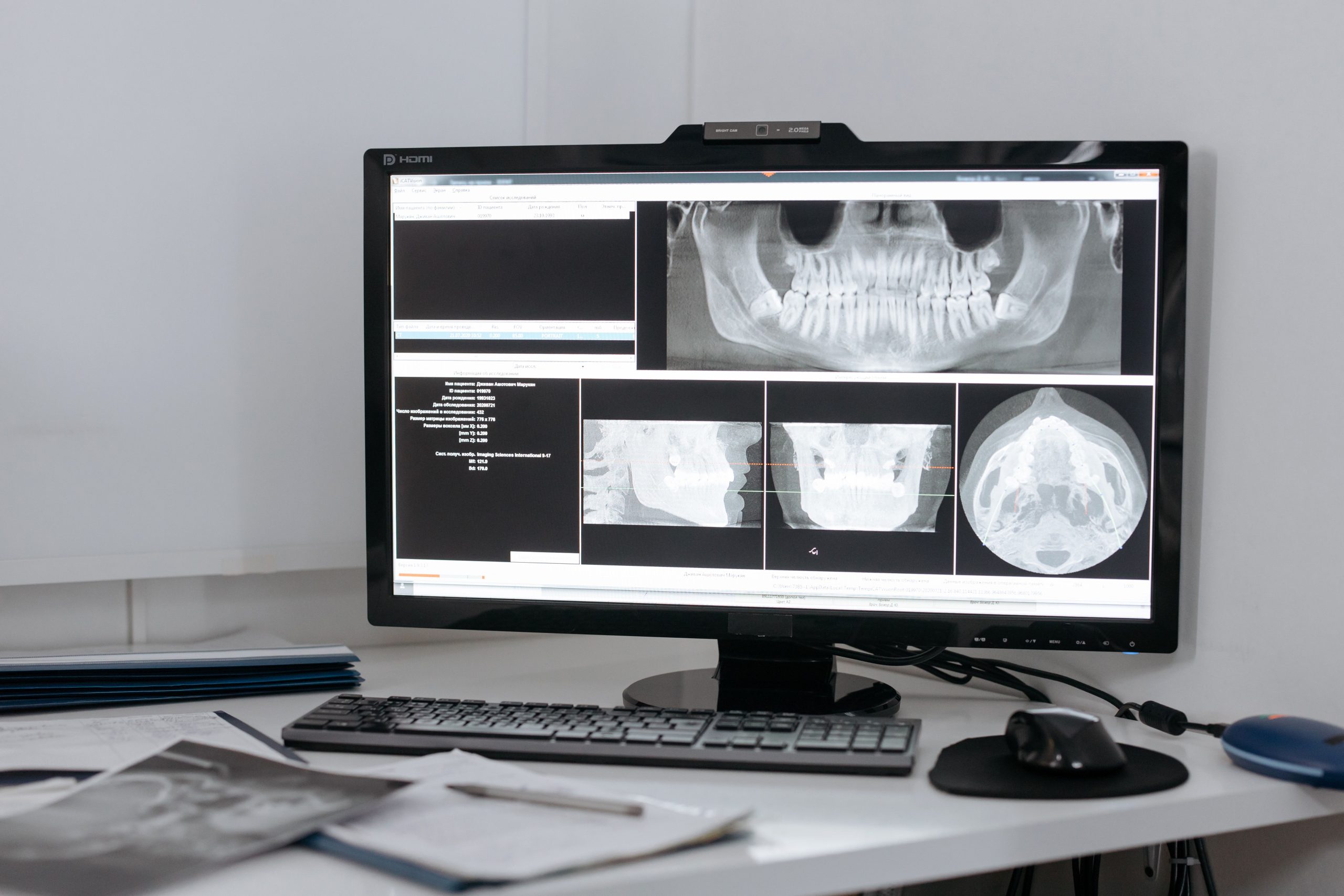
Clicking sounds from the jaw joint are often considered trivial. Many patients hear them while chewing or opening their mouths wide and simply choose to ignore them. Yet a click may be an early sign of displacement of the articular disc, a thin cartilage structure that helps the temporomandibular joint (TMJ) function smoothly. To determine what is actually happening inside the joint, dentists need accurate radiographic imaging rather than simple estimation.
The question is: Is a conventional panoramic radiograph sufficient, or is CBCT, which is more expensive and not always readily available, always necessary?
A study published in the Journal of Stomatology in 2023 provides a clear answer, and the findings are important for everyday clinical practice.
When Joint Space Becomes the Key to Diagnosis
The joint space is a radiolucent area, appearing dark on radiographic images, located between the mandibular condyle and the glenoid fossa. The distance in this area is not merely a number. It reflects the position of the condyle within the fossa, and changes in this distance may indicate abnormalities of the TMJ.
In this study, drg. Silviana Farrah Diba, Sp. RKG. Subsp. RP (K), together with a research team from the Department of Dentomaxillofacial Radiology, Faculty of Dentistry, Universitas Gadjah Mada, measured three joint-space parameters: anterior joint space (AJS), superior joint space (SJS), and posterior joint space (PJS). Measurements were performed in 34 patients aged 20–55 years who experienced clicking in one or both TMJs, with the jaws in centric occlusion.
Each patient underwent two imaging modalities: panoramic TMJ imaging (PanTMJ) using a Vatech PaX-I Insight machine and CBCT using an Orthopantomograph Instrumentarium Dental P 300. Measurements followed the Ikeda and Kawamura protocol, with sagittal sections serving as the primary observation plane.
Of the 34 participants, 31 were women, with a mean age of 26 years. Most participants experienced unilateral clicking. A history of orthodontic treatment was the most common predisposing factor, followed by the habit of chewing on one side.
Different Numbers, Different Conclusions
Statistical analysis using the Mann-Whitney test showed significant differences between PanTMJ and CBCT measurements for all three joint-space parameters (p < 0.05). These were not minor differences that could simply be ignored but clinically meaningful discrepancies.
On CBCT, the mean AJS was 2.50 mm and the mean SJS was 2.69 mm, both of which were greater than the measurements obtained using PanTMJ. In contrast, the PJS measured on PanTMJ (2.38 mm) was greater than that measured on CBCT. This means that panoramic imaging tends to underestimate the anterior and superior joint spaces while overestimating the posterior joint space.
“The results of this study show that joint-space measurements on PanTMJ cannot be compared with CBCT. For evaluating the condyle-fossa space, CBCT is the most reliable method because it provides multiplanar views.”
Why can the measurements differ so substantially? One explanation lies in the anatomy. When measuring the posterior joint space, the glenoid fossa is visualized together with the cranial base. Superimposition with the mastoid air cells of the temporal bone makes it difficult to determine measurement points precisely. Geometric distortion and image magnification in panoramic imaging also introduce uncertainty, particularly when the patient's head is not positioned perfectly.
CBCT, on the other hand, allows three-dimensional reconstruction that can be sectioned according to the condylar axis. The result is considerably more accurate for quantitatively measuring the condyle-fossa distance.
Panoramic Imaging Is Still Useful, but It Has Limitations
These findings do not mean that panoramic radiographs are useless. Panoramic imaging remains a cornerstone of dental radiographic examination: it is relatively inexpensive, widely accessible, and involves a lower radiation dose. In Indonesia, CBCT remains concentrated in major cities and is considerably more expensive. For many patients in rural areas, panoramic imaging may be the only available option.
However, the study emphasizes one important point: joint-space measurements on TMJ panoramic images cannot replace CBCT for quantitatively determining condylar position. Panoramic imaging remains relevant as an initial screening tool for detecting obvious morphological changes in the condyle. However, when clinicians require precise measurements to determine whether the condyle has shifted anteriorly, superiorly, or posteriorly, CBCT is the more reliable option.
The study also acknowledged several limitations. The position of the articular disc was not confirmed using MRI, meaning that the observed clicking was based solely on the clinical criteria of the RDC/TMD. The participants were also not stratified by sex, even though several previous studies have suggested that joint-space dimensions tend to be greater in men than in women.
Looking ahead, an interesting question remains: if panoramic imaging cannot reliably measure joint space quantitatively, can condylar morphology observed on panoramic images provide other clinically useful information? Future studies integrating MRI, CBCT, and panoramic imaging within a single study design may provide a more comprehensive picture while paving the way for more efficient and affordable TMD diagnostic protocols.
A clicking jaw joint is not always painful. But behind that sound, a small structure may be struggling to maintain its position. And to see it clearly, using the right imaging modality makes all the difference.
Source DOI: DOI: https://doi.org/10.5114/jos.2023.133645
Authors: Anny Anggraini, drg. Achmad Zam Zam Aghasy, M.Kes. Photo: Pexels
Photo: Pexels