
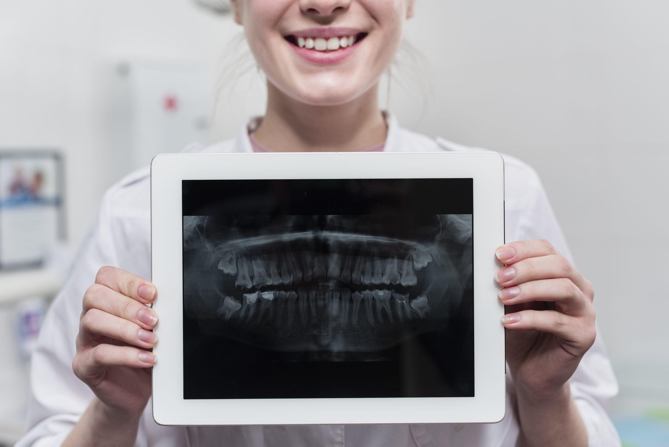
Imagine a postmenopausal woman visiting a dental clinic to have a cavity filled. Without realizing it, her periapical radiograph may contain information extending far beyond the condition of her tooth roots. This is where Dr. drg. Rini Widyaningrum, M.Biotech, saw an opportunity: the mandibular bone captured in a single radiograph could serve as a reflection of the condition of the patient's spine and hip.
This finding is not merely speculative. A study published in the International Journal of Electrical and Computer Engineering (IJECE), Volume 8, Issue 4, in 2018 demonstrated that analysis of periapical radiographic images could significantly correlate with bone mineral density in the lumbar spine and hip.
Two in Five Indonesians at Risk, but the Equipment Is Expensive
Osteoporosis is more than simply fragile bones. It is a systemic disease that increases the risk of fractures, reduces quality of life, and imposes substantial healthcare costs. The Indonesian Ministry of Health has reported that two out of five Indonesians are at risk of developing osteoporosis—a figure significant enough to make it a national health priority.
The problem lies in the diagnostic equipment. The current gold standard for measuring bone mineral density is Dual X-ray Absorptiometry (DEXA), which is recognized by the WHO. However, DEXA machines are expensive, unevenly distributed, and unavailable at every community health center or dental clinic. In contrast, dental X-ray equipment is available at nearly every dental healthcare facility in Indonesia, from major cities to smaller towns.
This is the gap that the study sought to address.
The Mandible as a Window into the Skeleton
Dr. drg. Rini Widyaningrum, M.Biotech, together with Sri Lestari from Universitas Respati Yogyakarta and Ferry Jie from Edith Cowan University, Australia, recruited 37 postmenopausal women as study participants. Each participant underwent two procedures: a DEXA scan at Dr. Sardjito General Hospital in Yogyakarta to measure bone mineral density in the hip and lumbar spine, and periapical radiography at Prof. Soedomo Dental Hospital, Faculty of Dentistry, UGM.
From the periapical radiographs, the research team extracted two parameters using ImageJ software and the Canny edge detection method: area parameter, defined as the proportion of detected trabecular area relative to the total area, and integrated density, calculated by multiplying the area by the mean pixel grayscale value. Both parameters reflect the condition of mandibular trabecular bone connectivity—the porous bone network that responds to systemic changes in bone mineral density.
The results were noteworthy. The area parameter showed a significant negative correlation with lumbar spine BMD (r = −0.371) and lumbar spine T-score (r = −0.383). In other words, the higher the area parameter value on the radiograph, the lower the bone density of the spine. Linear regression analysis further demonstrated that the area parameter could be used to predict lumbar spine T-score (F = 5.822, α < 0.05).
“The area parameter can be used as one of input parameters for computer-aided system of osteoporosis early detection by using periapical radiograph.” — Dr. drg. Rini Widyaningrum, M.Biotech, IJECE, 2018
Integrated density also showed a significant negative correlation with hip T-score (r = −0.332) and lumbar spine T-score (r = −0.377), although the association was not strong enough for it to serve as an independent predictor in the regression analysis.
Why the Lumbar Spine Rather Than the Hip?
One interesting finding was the stronger correlation with the lumbar spine compared with the hip. The explanation lies in bone composition: the lumbar spine consists of approximately 66% trabecular bone, whereas the hip contains approximately 25%. Because the mandibular trabecular bone analyzed in the periapical radiographs is also trabecular tissue, systemic changes in trabecular bone may be more readily reflected in parallel between the mandible and the spine.
As osteoporosis progresses, perforation of mandibular trabecular bone increases. This causes the area parameter value to rise because more “gaps” are detected by Canny edge detection. This pattern is correlated with a decline in lumbar spine T-score.
Although the correlations identified in the study were weak to moderate, the researchers emphasized that the findings represent a valid first step toward developing a computer-aided diagnosis (CAD) system for osteoporosis screening based on dental radiographs. An R-squared value of 14.6% for predicting lumbar spine T-score is relatively small, but it remains meaningful as one of multiple input parameters that could eventually be integrated into a more comprehensive CAD system.
A Small Step Toward Affordable Early Detection
The study has clear practical implications. If a CAD system based on periapical radiographs can be fully developed, even a dentist working in a small clinic could provide patients with an early warning of possible osteoporosis using routine dental X-rays. No DEXA machine costing hundreds of millions of rupiah would be required, nor would patients necessarily need immediate referral to a major hospital.
The study also represents one of the early bodies of literature specifically examining postmenopausal women from an Indonesian population, a group that has been underrepresented in similar studies dominated by Western populations.
A woman who visits a dental clinic simply to have a cavity filled may unknowingly be carrying important information about her own skeletal health. What is needed is a system intelligent enough to read it.
Source DOI: DOI: 10.11591/ijece.v8i4.pp2083-2090
Authors: Anny Anggraini; drg. Achmad Zam Zam Aghasy, M.Kes.
Photo: Freepik