
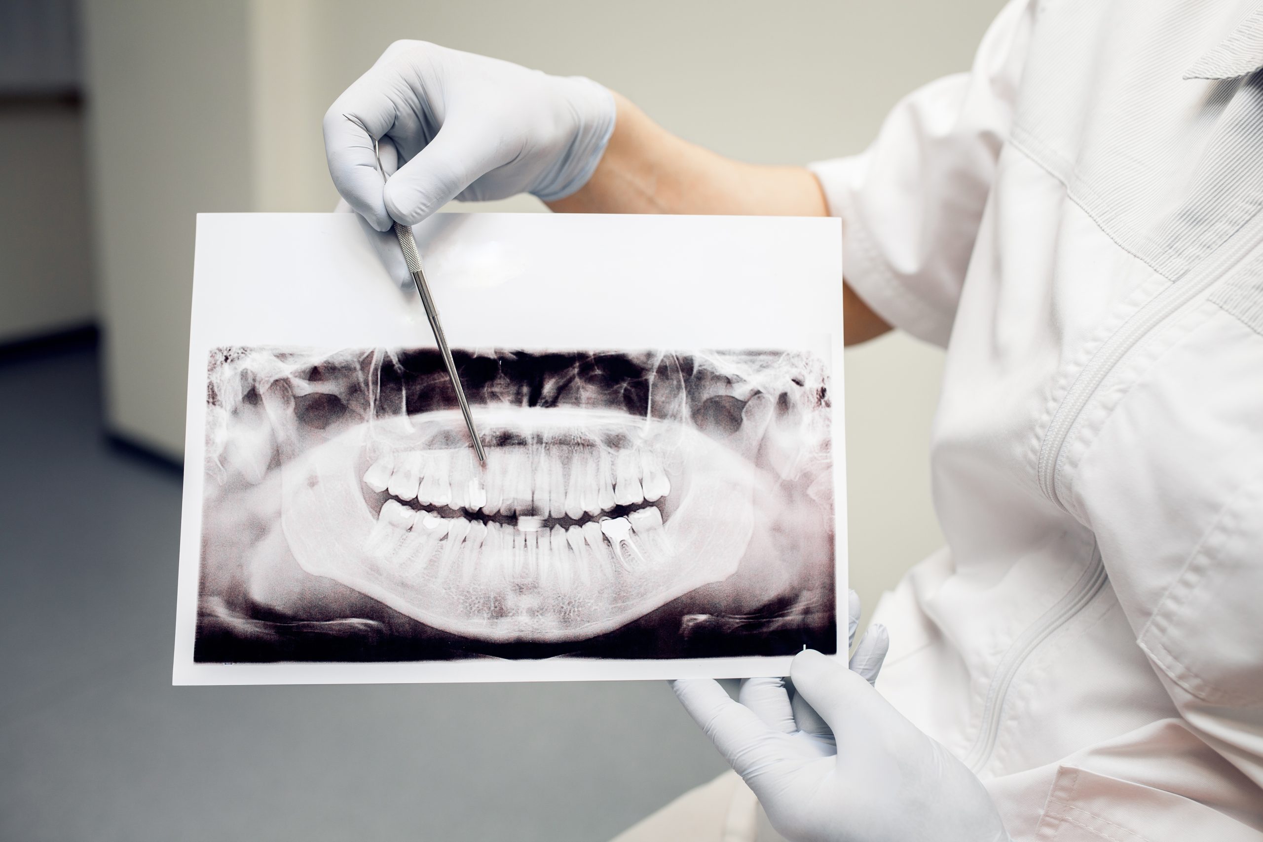
Every time a patient stands in front of a panoramic X-ray machine at a dental clinic, X-rays rotate around their head within seconds. The result is a comprehensive image of the entire jaw, helping dentists diagnose conditions affecting the teeth, bones, and even the sinuses. But behind this convenience lies a question that has not yet been fully answered: what actually happens to the cells inside a patient's mouth immediately after radiation exposure?
Dr. Ryna Dwi Yanuaryska, Ph.D., from the Department of Dentomaxillofacial Radiology, Faculty of Dentistry, Universitas Gadjah Mada, sought to answer this question directly at the molecular level. Published in the Journal of Dentistry Indonesia in 2018, the study was the first to use the comet assay to observe DNA damage in human buccal mucosal cells following panoramic radiographic exposure.
From Cheek Cells to a Fluorescence Microscope
The study involved 20 patients who underwent routine panoramic examinations at Prof. Soedomo Dental Hospital, Faculty of Dentistry, UGM. They were divided into two groups: one group had cheek-cell samples collected 30 minutes after panoramic radiography, while samples from the other group were collected 24 hours later. Samples were also collected from all participants before exposure to provide baseline measurements.
Buccal mucosal cells were selected for a reason. They can be collected easily by gently brushing the inside of the cheek with a small brush (cytobrush), making the procedure considerably more comfortable than obtaining lymphocytes from blood, which is invasive. After the cells were collected, the comet assay was performed: the cells were embedded in agarose gel, lysed, and then exposed to an electric field. Intact DNA moves slowly, whereas damaged DNA fragments migrate more rapidly, forming a characteristic “comet tail” under a fluorescence microscope.
The analysis was performed using OpenComet software, which automatically identifies comet structures and measures the percentage of DNA in the tail (tail DNA%), a highly sensitive parameter for detecting double-strand breaks (DSBs), in which both strands of the DNA molecule are broken.
Damage Appears, Then Disappears
The findings were both surprising and reassuring. In the group whose samples were collected 30 minutes after exposure, tail DNA% increased significantly (p=0.014). This indicates that radiation from panoramic X-rays indeed triggered DSBs in buccal mucosal cells within half an hour. However, in the group sampled 24 hours later, the difference was no longer detectable (p=0.29). The level of DNA damage had returned to its pre-exposure condition.
“DSBs can be induced in buccal mucosal cells 30 minutes after exposure to panoramic radiation, emphasizing the importance of the three principles of radiation protection: justification, optimization, and dose limitation.” — Dr. Ryna Dwi Yanuaryska, Ph.D.
The recovery observed within 24 hours was not coincidental. Human cells possess sophisticated DNA repair mechanisms. Once damage is detected, the MRN protein complex (Mre11-Rad50-Nbs1) actively recruits a series of repair proteins to the damaged site. Two major mechanisms involved are non-homologous end joining and homologous recombination, both of which can reconnect broken DNA strands.
Low Dose Does Not Mean Zero Risk
The radiation dose used in panoramic radiography is indeed far below the safety limits established by the International Commission on Radiological Protection (ICRP). Panoramic radiography uses approximately 2.7–38 microsieverts, while the stated annual permissible limit is 0.3 millisieverts. Nevertheless, the findings serve as a reminder that no radiation dose is completely free from potential biological effects.
The principle of radiation protection states that all radiation doses, regardless of magnitude, are considered potentially capable of producing effects. This concept is known as the linear no-threshold model, which forms the basis of global radiation safety regulations. Dr. Ryna's research provides concrete evidence that this principle is not merely a conservative theoretical assumption.
The findings also raise unanswered questions: which genes are involved in the DNA repair process in buccal mucosal cells following low-dose exposure? How quickly does the repair process actually begin? Further studies using narrower observation windows and gene-expression analyses are expected to provide answers.
Implications for Everyday Clinical Practice
Panoramic radiography is one of the most routine procedures in dental clinics. It is used to evaluate wisdom tooth development, plan dental implants, and detect jaw cysts, among other applications. Millions of people undergo the procedure worldwide each year. This study does not suggest that panoramic radiography is dangerous or should be avoided. Instead, it highlights something that can sometimes be overlooked amid routine clinical practice: every radiographic examination should have a clear clinical indication, use the lowest radiation dose that provides an adequate diagnostic image (as low as reasonably achievable), and employ radiation protection measures such as lead aprons when appropriate.
DNA damage that resolves on its own within 24 hours may not sound particularly concerning. However, over the long term, especially for patients who undergo numerous radiographic procedures throughout their lives, cumulative radiation exposure remains a consideration that should not simply be disregarded.
Source DOI: doi: 10.14693/jdi.v25i1.1124
Authors: Anny Anggraini, drg. Achmad Zam Zam Aghasy, M.Kes. Photo: Pexels
Photo: Freepik