
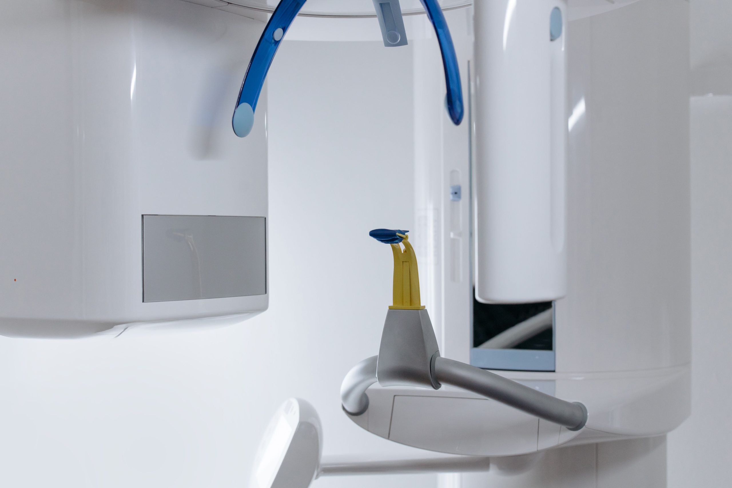
Maxillary expansion in children has traditionally been discussed primarily in the context of dental crowding or crossbite. However, a recent meta-analysis from the Faculty of Dentistry, Universitas Gadjah Mada (UGM), has revealed something more: this orthodontic procedure may also significantly increase the dimensions of the upper airway, including the nasal cavity, nasopharynx, and oropharynx.
Published in Dentistry Review (Elsevier, 2026), the study was a collaboration between the UGM orthodontic residency team, drg. Anrizandy Narwidina, MDSc, Sp.KGA, PhD, from the Department of Pediatric Dentistry, and Prof. Ananto Ali Alhasyimi from the Department of Orthodontics. The team analyzed 13 studies from around the world, all of which used cone-beam computed tomography (CBCT) for three-dimensional assessment.
A Narrow Jaw, Impaired Breathing
Maxillary deficiency in growing children is not simply a matter of facial aesthetics or dental alignment. Anatomically, the maxilla forms the floor of the nasal cavity and influences the nasopharyngeal and oropharyngeal spaces. When the upper jaw is too narrow or positioned too far posteriorly, surrounding structures may also be affected, including the airway through which a child breathes.
Maxillary expansion, particularly rapid maxillary expansion (RME), works by widening the upper dental arch using an expansion appliance. Its effects on respiratory function have long been debated. Some studies have reported increases in airway volume, whereas others have found no significant changes. These conflicting findings prompted the UGM research team to conduct an evidence-based systematic synthesis.
The Numbers Speak
Of the 1,370 articles identified through PubMed, ScienceDirect, and several other databases through May 2026, the researchers ultimately selected 13 studies for qualitative analysis and 12 for quantitative meta-analysis. All studies used CBCT rather than two-dimensional radiography, which is susceptible to distortion and superimposition.
The results were fairly clear. Maxillary expansion was associated with significant increases in three airway regions: the nasal cavity showed the largest effect (standardized mean difference/SMD = 0.91; p < 0.001), followed by nasopharyngeal volume (SMD = 0.71; p < 0.001) and oropharyngeal volume (SMD = 0.54; p < 0.001).
“Nasal cavity volume showed the largest pooled effect; however, substantial heterogeneity was observed for nasopharyngeal and nasal cavity outcomes.” — Amly et al., Dentistry Review, 2026
The greatest increase in nasal cavity volume makes anatomical sense: opening the midpalatal suture directly widens the floor of the nasal cavity. However, the high heterogeneity observed among tooth-borne appliances (I² = 88.46%) is a reminder that average values cannot be interpreted in isolation without considering differences in protocols and appliance types.
Not All Appliances Work the Same Way
One of the most clinically practical findings concerns the type of expansion appliance used. When the researchers stratified the data according to appliance type, notable differences emerged, particularly in nasopharyngeal volume.
Conventional tooth-borne appliances, which are widely used clinically, did not demonstrate a significant change in nasopharyngeal volume (SMD = 0.65; p > 0.05). In contrast, hybrid and miniscrew-assisted appliances produced a significant increase (SMD = 0.90; p < 0.001). This difference may be related to the mechanism of force transmission: miniscrew-based appliances transmit force more directly to the circummaxillary sutures and skeletal structures closer to the nasopharynx, whereas conventional RME produces a more pronounced dentoalveolar component.
For clinicians, the implication is important: the assumption that conventional expansion automatically enlarges the nasopharynx needs to be reconsidered.
Between Volume and Function: A Gap Yet to Be Bridged
This is where the scientific caution of the study becomes particularly important. The researchers clearly distinguished between anatomical changes measurable through CBCT and actual respiratory function.
An increase in nasal or nasopharyngeal volume on CBCT does not automatically mean that a child breathes better. There has been no confirmation through rhinomanometry, polysomnography, or the apnea-hypopnea index. Most of the studies analyzed also lacked untreated control groups, making it difficult to distinguish treatment effects from normal growth of the child's airway.
Another point worth noting is that almost all patients in these studies were between 7 and 16 years old, a period during which the airway naturally enlarges with growth. Follow-up periods ranging from 23 days to 15 months further complicate causal interpretation.
The researchers emphasized that controlled clinical trials using validated respiratory endpoints are still urgently needed before firm clinical recommendations can be made. CBCT volume, after all, is an anatomical surrogate rather than a direct reflection of function.
For children with narrow maxillae sitting in the orthodontic chair, these findings raise the possibility that the benefits of expansion may extend beyond a straighter smile. However, the extent to which those benefits translate into improved everyday breathing remains a question for the next generation of research.
Source DOI: https://doi.org/10.1016/j.dentre.2026.100433
Authors: Nanda Ayu, drg. Achmad Zam Zam Aghasy, M.Kes.
Photo: Pexels