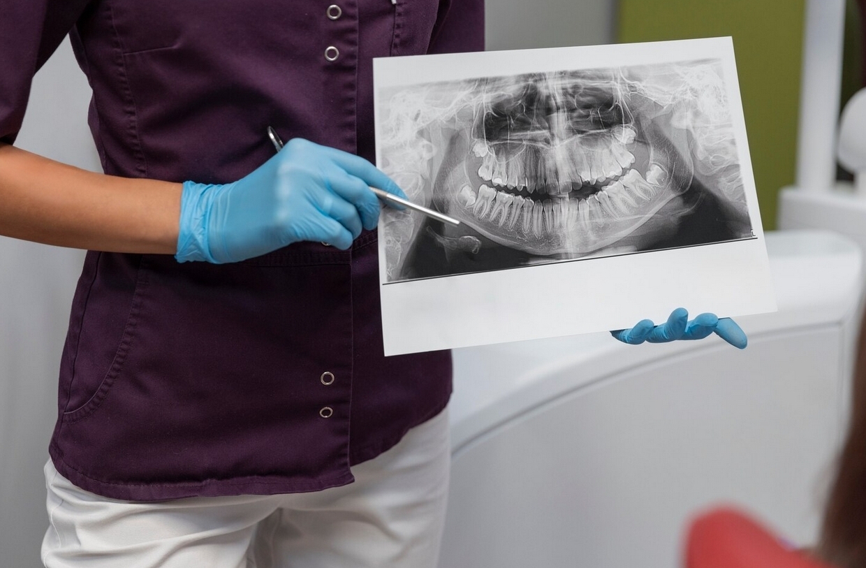
In dental radiology, particularly intraoral examinations such as periapical radiography, scoring systems are essential for assessing image quality. Radiographic scoring aims to determine whether images meet diagnostic standards (contrast, sharpness, exposure, positioning, artifacts) so that they are suitable for diagnosis and treatment planning.
A study conducted by Shoofia Aulia Rahma, a student of the Faculty of Dentistry, UGM, under the supervision of drg. Ryna Dwi Yanuaryska, Ph.D., and Dr. drg. Rini Widyaningrum, M.Biotech., entitled “The Correlation Between Radiograph Quality and Knowledge of Periapical Radiographic Examination Procedures (A Study on Co-Assistant Students of the Faculty of Dentistry, Universitas Gadjah Mada)”, examined the relationship between radiograph quality and operator knowledge of radiographic procedures.
Concepts and Principles of Dental Radiology Scoring
Key elements in radiographic scoring systems include:
- Image quality criteria: sharpness, contrast, proper exposure, sensor/film positioning, avoidance of artifacts, and appropriate illumination.
- Scoring scale: images are categorized into classes such as “unacceptable,” “acceptable/good,” and “excellent” based on fulfillment of quality criteria.
- Independent assessment: scores are typically assigned by trained observers or radiologists based on reference standards.
- Clinical purpose: high scores indicate diagnostically acceptable images for pathological interpretation and evaluation of roots, canals, and periapical structures.
Findings and Implications from UGM Research
The study found:
- A significant positive correlation between operator knowledge of periapical radiographic procedures and the quality of radiographs produced (p = 0.008; r = 0.477).
- This means that the higher the operator’s technical knowledge (positioning, exposure, technique), the better the image quality score.
- Images meeting more quality criteria were classified as “good” or “excellent,” while those failing to meet several criteria received lower scores.
Implications include:
- Scoring systems are effective quality indicators for determining whether images are suitable for diagnosis.
- Operator training and knowledge are critical, as image quality depends heavily on technical skills.
- Feedback for technical improvement can be obtained through routine scoring, allowing operators to analyze errors and improve future techniques.
Advantages and Limitations of Scoring Systems
Advantages:
- Provide objective benchmarks for image quality evaluation.
- Support standardization of radiographic quality among operators.
- Reduce the use of non-diagnostic images, thereby minimizing repeated radiation exposure.
Limitations:
- Observer subjectivity may vary; training and standardization are needed.
- Scoring focuses on technical aspects, not pathological interpretation.
- Images of “acceptable” quality may still be diagnostically usable in certain cases.
***
Dental radiology scoring systems are essential tools to ensure that radiographic images meet diagnostic standards. UGM research demonstrates that good image quality strongly depends on the operator’s technical knowledge, as reflected in radiograph quality scores. By adopting scoring systems, dental radiology practice can improve image quality, reduce repeat errors, and support more accurate diagnosis.
References
SHOOFIA AULIA RAHMA, drg. Ryna Dwi Yanuaryska, Ph.D; Dr. drg. Rini Widyaningrum, M.Biotech., The Correlation Between Radiograph Quality and Knowledge of Periapical Radiographic Examination Procedures (A Study on Co-Assistant Students of the Faculty of Dentistry, Universitas Gadjah Mada), https://etd.repository.ugm.ac.id/penelitian/detail/222277
Author: Rizky B. Hendrawan | Photo: Freepik