
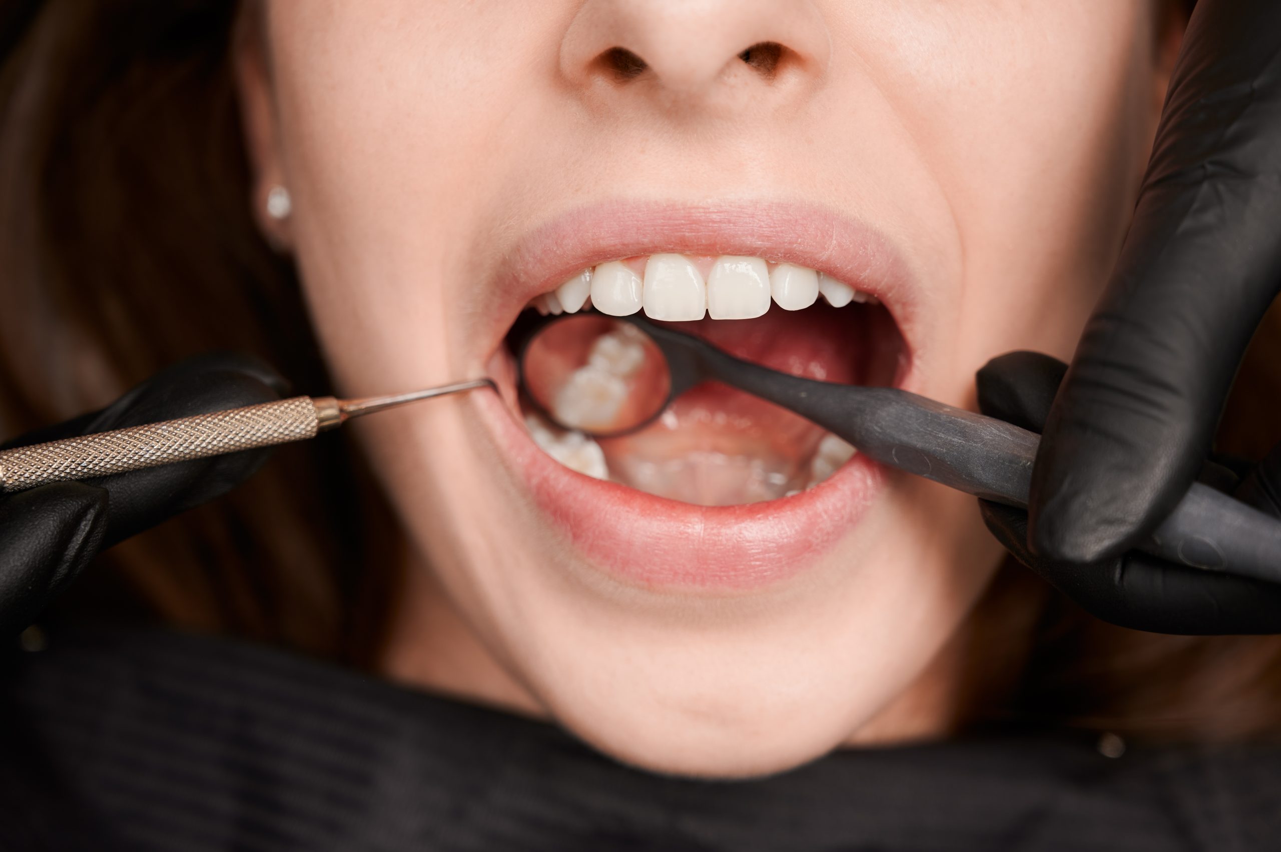
A lecturer and researcher from the Department of Conservative Dentistry, Faculty of Dentistry, Universitas Gadjah Mada (FKG UGM), drg. Margareta Rinastiti, M.Kes., Sp.KG(K)., Ph.D., together with a research team from UGM and the University of Groningen, the Netherlands, has successfully found a scientific solution to a clinical dilemma frequently faced by dental practitioners. This collaborative research demonstrated that the repair procedure (relayering) for deteriorating resin composite fillings can achieve very strong results, provided that the surface preparation method is properly performed using a silica coating technique (silica coating).
The comprehensive findings, which have been successfully published in the reputable international journal Clinical Oral Investigations, provide a clear answer for minimizing the need to completely remove dental fillings. This experiment makes an important contribution to supporting the philosophy of modern dentistry, which prioritizes the preservation of healthy natural tooth tissue (minimally invasive dentistry) to improve the quality of oral healthcare for the community.
To date, when a patient’s resin composite filling ages and begins to show deterioration in the oral cavity, such as discoloration, marginal leakage, wear, or cracking, the most common procedure is to replace the entire filling. However, most fillings that are replaced are actually still in clinically good condition. Such complete replacement forces dentists to remove additional healthy tooth tissue, which in the long term can weaken the sound tooth structure and risk damaging the pulp, the living tissue inside the tooth.
As a more conservative solution, repairing the filling by adding a new layer over the old filling (relayering) has become the best alternative. However, the effectiveness of this method has been debated because the bond strength between aged fillings and new material tends to decrease drastically, by 25 to 80 percent compared with non-aged fillings. To address this problem, drg. Margareta Rinastiti and her team conducted a laboratory experiment testing four commonly used resin composites by simulating real-world aging conditions through temperature fluctuations (thermocycling), immersion in water at body temperature, and exposure to citric acid.
After undergoing artificial aging, two surface preparation methods were applied before the new material was placed. First, the application of an intermediate adhesive resin (intermediate adhesive resin, IAR). Second, a more complex method involving tribochemical silica coating using 30-micrometer alumina particles coated with silica, followed by the application of silane and then intermediate adhesive resin, referred to as the SC (silica coating) method.
The research results showed a consistent pattern, although not as straightforward as expected. In general, the SC method produced higher bond strengths than IAR, particularly after aging through water storage. Under these conditions, three of the four types of composite even achieved bond strengths exceeding those of the non-aged control, an unexpected finding.
“SC-application led to higher bond strengths than IAR-application after thermocycling and water storage. In addition, SC-application led to more cohesive failures than after IAR-application, regardless the aging method.” Rinastiti et al., Clinical Oral Investigations, 2011
Beyond the bond strength values, the significance of this research can be seen in the analysis of failure patterns under a microscope. The SC method was shown to produce cohesive failures, meaning that the part that fractured under testing pressure was not the adhesive bond at the interface between the old and new fillings, but the filling material itself. This serves as a valid indicator that the bond formed by the silica method was actually stronger than the underlying composite material. In contrast, the conventional adhesive method (IAR) more frequently produced adhesive failures, meaning that the bond detached precisely at the interface between the old and new fillings, indicating that the bond itself had become the weak point.
Nevertheless, the researchers noted that after aging through thermocycling and aggressive citric acid immersion, even the SC method was unable to restore bond strength to the initial level. Surfaces that had been excessively degraded by extreme temperature fluctuations and acid attack made it difficult for silica particles to adhere because the surface was already dominated by exposed filler particles rather than the softer resin matrix.
Clinically, these findings have practical implications that can be directly applied in dental practice. The results provide a stronger scientific basis for determining when and how repairs should be performed precisely. For fillings with mild to moderate degradation, for example due to prolonged exposure to water and saliva, the SC method is worthy of being the primary choice because it has consistently been shown to be more reliable, although the procedure is longer than simply applying conventional adhesive resin. Meanwhile, for fillings that have undergone severe damage due to thermal stress or prolonged acid exposure, complete replacement may still need to be considered.
Through valid scientific data, UGM continues to promote research downstreaming that respects patients’ biological structures. Every millimeter of tooth tissue that can be preserved through appropriate clinical decisions is a long-term investment in patients’ dental health. The research by drg. Margareta Rinastiti and her team reminds us that the success of dental treatment is not measured solely by the speed or convenience of a procedure, but by choosing the medical approach that best respects the tooth itself.
Author: drg. Achmad Zam Zam Aghasy, M.Kes., Annisa Dwi Noviyanti
Photo: FreePik
Source DOI: 10.1007/s00784-010-0426-6