
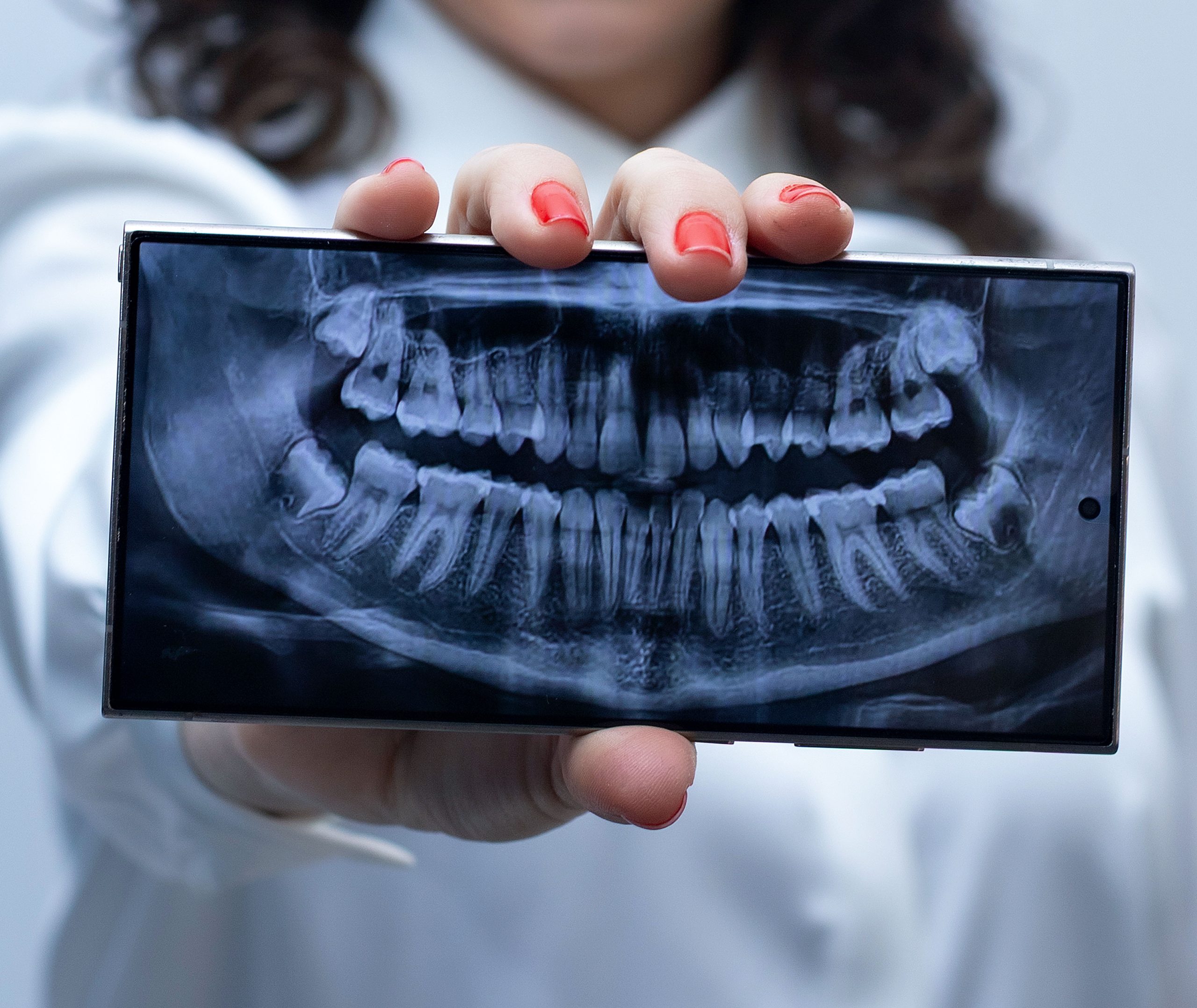
A dental panoramic image contains an extraordinary amount of information. Within a single 2,880 × 1,504-pixel image are 32 teeth, the jawbones, sinuses, joints, and numerous overlapping anatomical structures. Among all of these, clinicians must identify a small but potentially consequential abnormality: an unerupted canine, clinically known as an impacted canine. This task requires a trained eye, time, and a high level of concentration. Now, an artificial intelligence system developed by researchers from KU Leuven, Belgium, has demonstrated near-perfect accuracy in performing this task.
When Canines Hide Behind Bone
The canine, or eye tooth, has the longest root among the teeth in the oral cavity. Its strategic position helps shape the dental arch and plays an important role in both mastication and facial aesthetics. When a canine fails to erupt and remains trapped within the jawbone, the condition is known as impaction. Impacted canines are not rare: they are the second most common type of impacted tooth after third molars. If left untreated, they can cause root resorption of adjacent teeth, cyst formation, and complex occlusal problems.
Detecting them on panoramic images, however, is not easy. Large image dimensions, considerable variation in the position of impacted teeth, and the density of surrounding anatomical structures make manual identification time-consuming and susceptible to interpretive errors. This was the starting point for research involving drg. Rellyca Sola Gracea, Sp.RKG. Subsp.RDP (K), a faculty member of the Faculty of Dentistry at Universitas Gadjah Mada who is currently affiliated with the OMFS-IMPATH Research Group at KU Leuven, together with researchers Sirin Guner Onur, Soroush Baseri Saadi, and Reinhilde Jacobs.
Two Steps, One Goal
The research team designed a two-stage deep learning system with an elegant approach. Rather than asking the algorithm to analyze an enormous full-size panoramic image immediately, the system works much like a clinician who first scans the entire image and then zooms in on suspicious areas for closer examination.
The first stage uses a YOLOv8-based detection model to identify the location of impacted canines in the full image, marking them with a bounding box. Once the location is identified, the second stage takes over: a segmentation model analyzes the cropped and resized region and precisely traces the contour of the impacted tooth pixel by pixel.
The dataset consisted of 143 high-resolution panoramic images, with 114 images used for training and validation and 29 for testing. The entire training process employed fivefold cross-validation, a method designed to ensure that system performance is not dependent on a single subset of the data.
The results exceeded expectations. The detection model achieved a precision of 0.941 and a recall of 0.887. The segmentation model performed even better, with a precision of 0.956 and a recall of 0.905. Most impressive was the mean average precision (mAP) at an IoU threshold of 0.50, which reached 0.986 for both tasks. On a scale from 0 to 1, this figure is remarkably close to perfect.
“The proposed two-stage deep learning pipeline demonstrates high accuracy in detecting and segmenting impacted canines, offering a reliable automated tool for clinical diagnosis and treatment planning in dental practice.” — drg. Rellyca Sola Gracea, Sp.RKG. Subsp. RDP (K), et al., Journal of Dentistry, 2026
From Pixels to the Dental Chair
What do these numbers mean for patients sitting in the dental chair? In practical terms, the system could potentially reduce the time required to interpret radiographs, lower the risk of missed diagnoses, and provide more informative visualizations for both clinicians and patients. The system's output is not limited to numerical data. It also generates an image with the contour of the impacted tooth superimposed directly onto the original panoramic image, making it easier for dentists to plan orthodontic or surgical treatment.
The contribution of drg. Rellyca, who is also a specialist in Oral Radiology with a subspecialization in Pediatric Dentomaxillofacial Radiology from FKG UGM, adds an important clinical dimension to the research: the perspective of a practitioner who understands the everyday challenges of radiographic diagnosis. The study was published in Journal of Dentistry (2026), one of the world's leading dental journals.
Of course, the system is not yet perfect. A dataset of 143 images is relatively small by deep learning training standards, and its performance in more diverse patient populations requires further evaluation. Nevertheless, as a proof of concept, the system provides a strong foundation: artificial intelligence is not merely an administrative aid in the dental clinic but an increasingly viable diagnostic partner.
At some point in the not-too-distant future, a newly captured panoramic image may arrive with automatic annotations already in place, highlighting every abnormality before the dentist even touches the mouse. What remains beyond the reach of any algorithm, however, is clinical decision-making, empathy toward patients, and the comprehensive judgment of a clinician.
Source DOI: https://doi.org/10.1016/j.jdent.2026.106463
Authors: Anny Anggraini; drg. Achmad Zam Zam Aghasy, M.Kes.
Photo: Pexels