
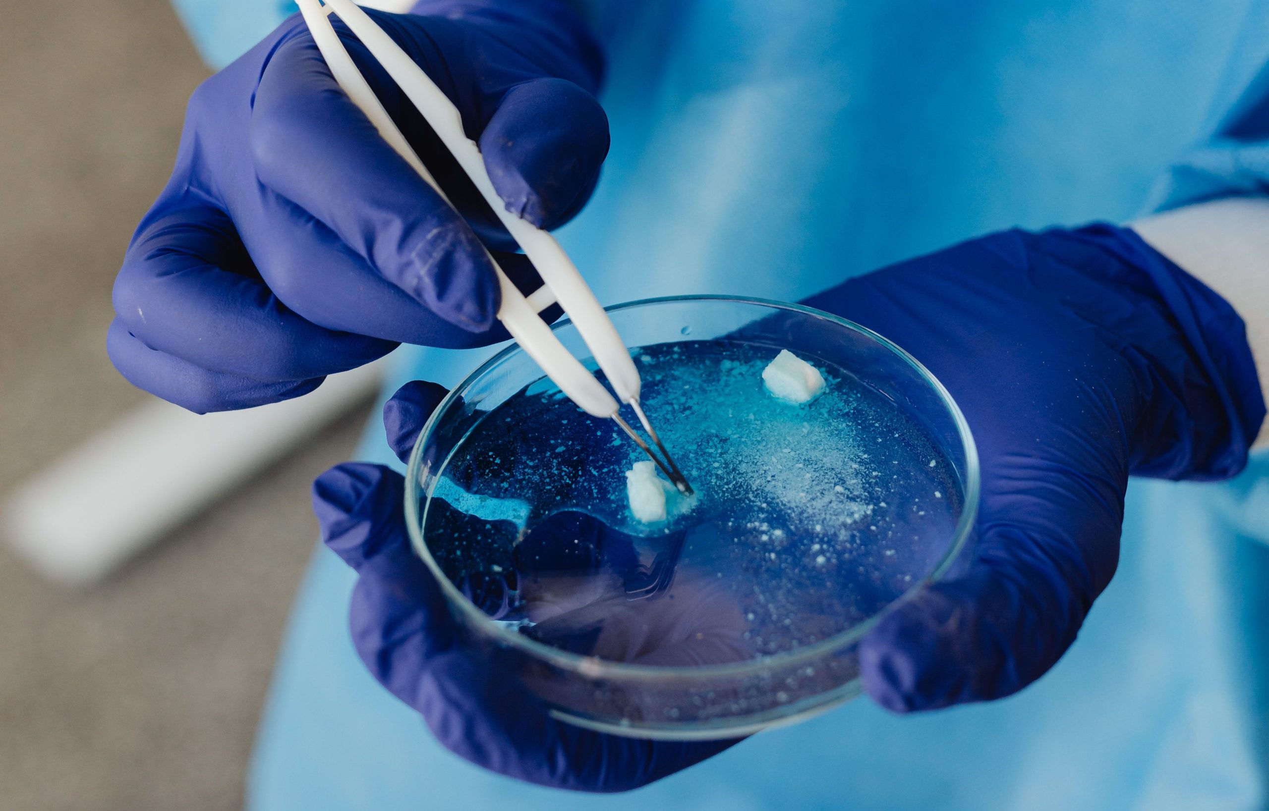
Until now, the sensation of pain when a tooth is exposed to cold or heat has generally been regarded simply as a pain signal. Yet something much more complex is happening beneath the surface: fluid within the dentinal tubules moves, exerting pressure on odontoblasts and triggering a biological response that goes far beyond pain. The question is, how is this mechanical pressure ultimately translated into the formation of new dentin?
A study published in Scientific Reports (2026) provides a more concrete answer than ever before. A collaborative study involving Tokushima University, the Institute of Science Tokyo, and Universitas Gadjah Mada successfully mapped the molecular signalling pathway linking hydrostatic pressure to odontoblast differentiation through three key players: PIEZO1, RUNX2, and WNT16. One of the researchers involved in data acquisition and analysis was drg. Anrizandy Narwidina, MDSc, Sp.KGA, Ph.D., a lecturer in the Department of Pediatric Dentistry at Universitas Gadjah Mada.
Stem Cells from Naturally Shed Baby Teeth
The biological material used in this study is not something commonly encountered in general biology laboratories: stem cells derived from naturally shed human baby teeth, known as SHED (Stem Cells from Human Exfoliated Deciduous Teeth). These cells were collected from 24 healthy children aged 6 to 13 years who received treatment at Tokushima University Hospital. The teeth used were caries-free, and all procedures were performed with written consent from both the parents and the children involved.
SHED are not merely “leftovers” from teeth that are no longer needed. These cells possess strong differentiation potential, including the ability to develop into odontoblasts, the cells responsible for forming dentin. The researchers then subjected the cells to a hydrostatic pressure of 0.5 kPa, mimicking the physiological pressure that naturally occurs within dentinal tubules.
The results showed a significant increase in the expression of odontoblast marker genes, namely PANX3 and DSPP. Mineralisation, measured using Alizarin Red S staining, also appeared denser and more extensive.
When Ion Channels Become the Gateway
This is where PIEZO1 enters the story. This mechanosensitive ion channel is known as a pressure sensor at the cell membrane. When hydrostatic pressure is applied, PIEZO1 opens, allowing calcium to enter the cell and triggering a downstream signalling cascade.
To establish its central role, the researchers used siRNA to “silence” the PIEZO1 gene. The results were striking: the mineralisation that had previously increased in response to hydrostatic pressure almost completely disappeared. The expression of PANX3 and DSPP also declined sharply. This demonstrated that PIEZO1 is not merely a supporting player, but rather a critical gateway that must be activated for mechanical pressure to be translated into a biological response.
However, PIEZO1 does not act alone. Its activation promotes the nuclear translocation of RUNX2, a transcription factor long recognised as a key regulator of odontoblast differentiation. Once in the nucleus, RUNX2 does something that had not previously been confirmed in this context: it directly binds to the promoter of the WNT16 gene and activates its transcription.
Direct Evidence of the Molecular Cascade
To establish the relationship between RUNX2 and WNT16, the researchers used two complementary approaches. First, a luciferase reporter assay in HEK293 cells showed that RUNX2 overexpression increased WNT16 promoter activity by up to 5.6-fold at the highest dose. Second, a HaloChIP assay confirmed that RUNX2 physically binds to a binding motif within the WNT16 promoter, with approximately seven-fold enrichment compared with the control.
“These findings identify PIEZO1–RUNX2–WNT16 signalling as a previously unrecognised pathway linking mechanical stimulation to odontoblast differentiation in SHED,” the researchers wrote in their paper.
Interestingly, of the two known human WNT16 isoforms, only WNT16b was detected in SHED. This confirms that this isoform is the functional target of the PIEZO1–RUNX2 pathway during odontogenesis.
When WNT16 expression was silenced using siRNA, hydrostatic-pressure-induced mineralisation decreased significantly, although it did not disappear completely. This suggests that WNT16 is a central player, but that other parallel pathways may also contribute, most likely through YAP/TAZ, another mechanosensitive pathway previously associated with PIEZO1.
From the Laboratory Bench to Future Clinical Applications
This discovery opens the door to new approaches in pulp therapy and dentin tissue regeneration. At present, the treatment of dentin damage still relies heavily on conventional restorative materials. However, if the PIEZO1–RUNX2–WNT16 pathway can be therapeutically manipulated—for example, through pharmacological activation of PIEZO1 or local delivery of WNT16 controlled stimulation of secondary and tertiary dentin formation could become a realistic possibility.
The researchers themselves acknowledge that the study is still based on a loss-of-function approach. Further experiments, such as gain-of-function studies or rescue experiments restoring WNT16 expression in PIEZO1-deficient cells, are still needed. Nevertheless, the molecular map now available is sufficient to envision a future in which injured teeth could be assisted in rebuilding themselves, with a little help from mechanisms already present within them.
A baby tooth naturally shed from the mouth of an eight-year-old child, it seems, still has much to teach the field of dentistry.
Source DOI: https://doi.org/10.1038/s41598-026-46415-y
Authors: Nanda Ayu, drg. Achmad Zam Zam Aghasy, M.Kes.
Photo: Pexels